![]()
Computer Assisted Analysis of 3D MRA Images
Introduction
Atherosclerosis
is the principal acquired affection of the vascular wall. It is one of the most
important problems of public health and is the number one cause of death among
people older than 60 years in the western countries. Its major complication is
the arterial stenosis, which is characterized by the
thickening of the artery wall and by the narrowing of its lumen. Quantitative
characterization of stenosis is of major interest in
medical vascular imaging. The development of image analysis techniques for
objective and precise quantitative analysis of 3D data is necessary to obtain
reliable and reproducible results.
We
deal with image processing applied to three-dimensional (3D) analysis of
vascular morphology in magnetic resonance angiography (MRA) images. The main
goal of our work is to develop a fast and reliable method for stenosis quantification. It consist
in three main steps:
·
Vessel axis
extraction by an expansible skeleton
method
·
Vessel boundaries
detection in the planes locally orthogonal to
the centerline using an improved active contour.
·
Quantification of the stenosis degree based on measurements
of the resulting contours
This work constituted my Ph.D. project and was carried out in the
framework of the dynamic imaging scientific topic and the vessels medical
project of CREATIS lab. It was supported by CARENA S.A and it was in the scope
of the scientific topics of the GDR-PRC ISIS research group of the
![]()
|
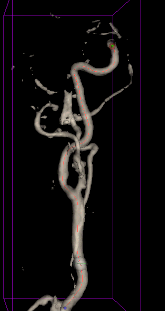
The
vessel axis extraction is achieved by an expansible skeleton method. It is
based on a tracking strategy, which begins from the computed starting point within
the vessel, and then iteratively estimates the subsequent axis points (at
each iteration, a new point is added to the model). Point generation is a
two-step procedure. First, a prediction of the new point position is
obtained, based on the vessel local orientation at the current point. The
vessel local orientation is estimated by inertia moment minimization for a
small volume centered on the current point. This predicted position is then
corrected under the influence of image forces and shape constraints.
|
|
![]()
|
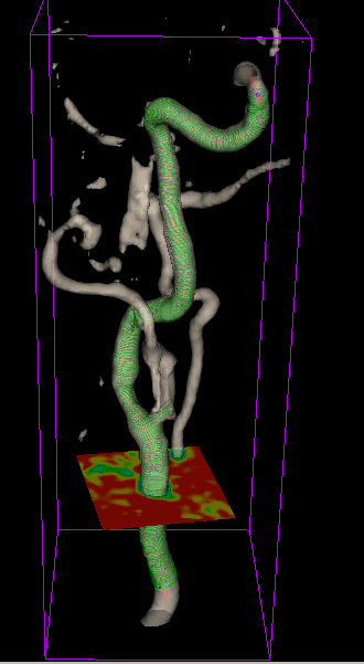
Vessel boundaries detection For the detection of vessel lumen boundaries in the planes locally
orthogonal to the vessel axis, we use a deformable model: an active contour.
The use of a balloon force together with a new numerical implementation
permits an initialization with a single pixel, which is automatically defined
by the intersection between the vessel axis and the orthogonal image plane. |
|
![]()
|
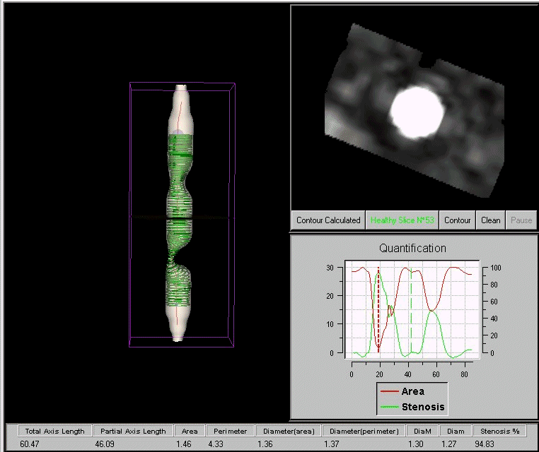
Quantification Vessel contours detection in the planes locally orthogonal to the
centerline results in a stack of 2D contours along the vessel, allowing quantitative
cross-section measurements. Furthermore, thus obtained outline can be
visualized by means of a triangulation-based rendering technique. |
|
![]()