F. Peyrin
Purpose and context
Although less studied than trabecular bone, cortical bone also plays an important role in osteoporosis : it is generally the site where the fractures are initiated. The purpose of this work was to quantify the 3D micro-porosity of cortical bone from synchrotron micro-CT.
Methods
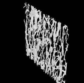
3D images of cortical bone were obtained from synchrotron micro-CT with a voxel size of 10 or 5 µm. Cortical porosity which can be obtained from the negative of the image, constitutes a very dense network, highly orientated. Quantitative parameters of the cortical bone porosity network were extracted. .
Results
The initial study on this topic was performed in collaboration with V. Bousson and C. Bergot (Hospital Lariboisière, Paris). Femoral head samples were acquired at 10 µm from elderly patients. We showed, for the first time, the possibility of obtaining qualitative and quantitative data on the three-dimensional organization of the Haversian network
|
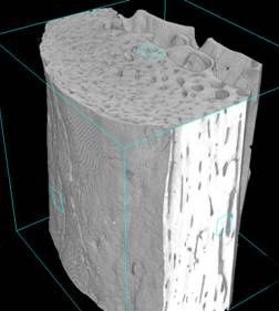
(a) |
(b) |
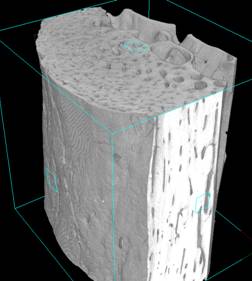
3D SR micro-CT reconstructed image of the inferior femoral neck cortex in humans. a) volume of the proximal region of the neck (near the head) b) illustration of the Haversian system (thicknes:300 µm)
In a second study, performed with the LIP (Laboratoire d’Imagerie Paramétrique, UMR CNRS 7623, Paris, P. Laugier), samples of cortical bone taken from human radiuses or femoral necks were imaged in order to explain ultrasound measurements. Images acquired at 10 µm were used to relate the ultrasound parameters measured in axial transmission on the same samples with the mineralization and cortical porosity
Perspectives include the automatic segmentation of remodeling zone from 3D synchrotron micro-CT images of cortical bone. Although such regions are visible, their contrast is low and they are corrupted by noise and ring artifacts, thus a specific segmentation method is required. This segmentation could enable to quantify new parameters of bone metabolism.
Collaborations and acknowledgements
This work was performed with Hospital Lariboisière, CNRS UMR 7052, Paris, LIP, CNRS UMR 7623 ,Paris, and Q-BAM Group, University of Halle-Wittenberg, Germany.D R.O., PEYRIN F., LAUGIER P., Derivation of elastic stiffness from site-matched mineral density and acoustic impedance maps, Physics Medicine and Biology, 2006, vol 51, n° 3, pp. 747-758.