(E. Brusseau, V. Detti)
In collaboration with the Hospices Civils de Lyon, Lyon, France.
The 2D strain estimation method we have developed is currently assessed at the radiology department of the Hôpital de la Croix-Rousse with patients suffering from breast lesions. The objective is to analyze and assess the complementary information revealed by imaging tissue deformation over compression.
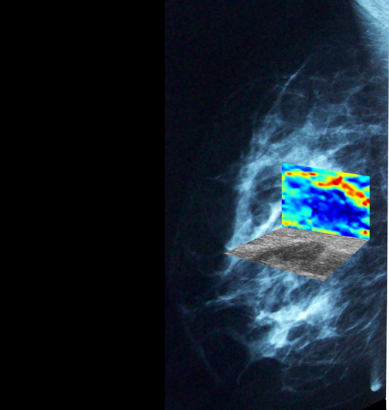
An illustration of elastography results with benign and malignant lesions is presented below.

Left : B-mode images, right : elastograms (axial strain)
We have observed that fibroadenomas tend to be stiffer than the surrounding tissues with similarities in size, shape and margin between elastograms and B-mode images. Carcinomas appear stiffer than the surrounding tissues, but the stiffer region is frequently much larger in the elastogram than the corresponding lesion in the sonogram, and with a different overall shape.
More information is available in the following references