Master Internship
Coherence imaging with Deep Learning
The heart is a complex organ that performs the essential function of circulating blood in the human body. This function is essential to life and heart disease remains an extremely important cause of death in industrialized countries. The development of diagnostic tools or therapeutic methods requires a detailed understanding of the physiology of the heart: motion / deformation of the muscle, hemodynamic in the various cavities, electrical activation, etc. Moreover, since the heart consists of muscle fibres, it also seems very relevant to try to image the local fibrous structure of the tissue as finely as possible in order to establish a link between this local structure and heart function and more generally with the development of the various pathologies.
Based on MRI imaging of the free diffusion of water in tissue, CREATIS is one of the world leaders in cardiac fibre imaging. This type of imaging is made very complex especially because of the rapid and significant movement of the heart during MRI acquisition. In addition, thanks to the emergence of ultra-fast ultrasound imaging by plane wave, a first technique for imaging tissue structure by ultrasound has recently been developed [1-2]. Ultrasound has a significant number of advantages over MRI, including its much lower cost, its portability and, for our application, its high acquisition speed, particularly in ultra-fast imaging.
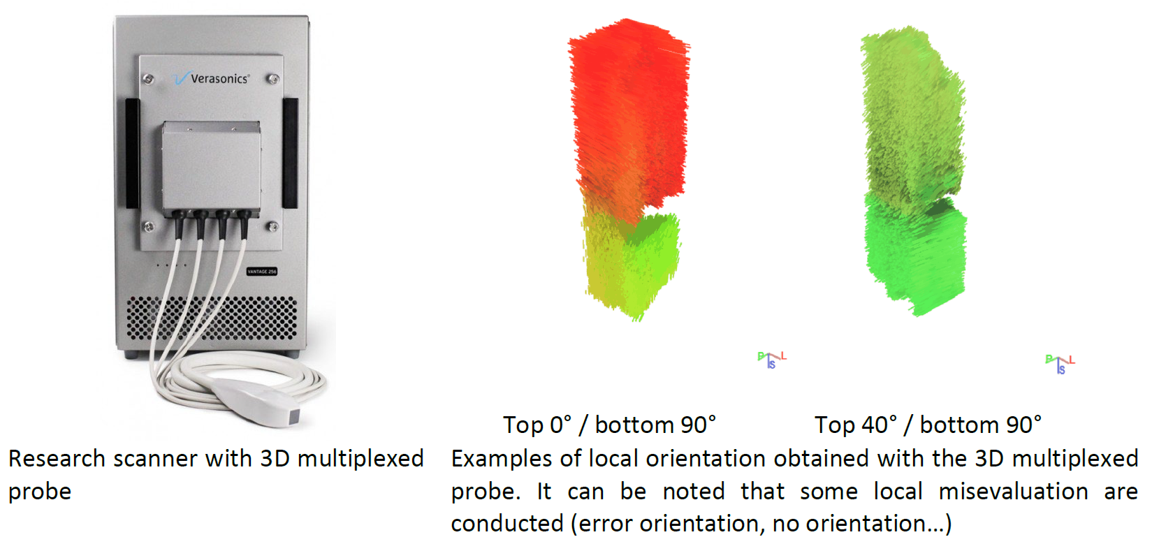
As part of the work carried out in the laboratory [3], the technique was developed and validated on various experimental environments. To carry out the acquisitions, the synchronization of 4 Verasonics Vantage 256 devices is required. This procedure is very time-consuming to implement, synchronization is capricious and the handling time is greatly reduced due to the rare availability of the 4 Verasonics systems. To alleviate this, a new multiplexed 3D ultrasonic probe (256, 512 or 1024 elements) was acquired, and allow, with some adaptations, to implement the imaging technique we have proposed from a single or two Verasonics systems. However, even with such improvement, the coherence technique did not perform as well as it was expected.
The purpose of this internship is to propose a deep learning strategy to evaluate the local tissue orientation in ultrasound imaging. To conduct such objective, the intern will have to generate several simulation dataset based on synthetic model of the Lab [4] coupled with experimental dataset previously acquired (or to acquire if the settings have changed). The arrangement of these numerous 3D raw signal dataset have to be conducted before exploit in the network identified by the intern. Depending on the advancement of the project, the potential utilisation on the network directly on the US scanner must be performed. It must be noted that no difficulty is present in this subject around the database generation and legal aspects.
The objectives of this internship are
Formalized the database for local orientation estimation (simulations and experiments)
Propose a deep learning strategy that exploit such database
Evaluate the requirement/potential for transferring this technique on the PiLoT platform
Depending on the results and following this initial work, a continuation of the thesis will be envisaged.
Profile/Skills: Student from a top engineering school (generalist or EEA profile) Image and signal processing, ultrasound imaging, deep learing, mathematics, etc.
Start and duration of the course: February/March 2022 for a duration of 6 months.
How to apply
Send CV + cover letter + M1/M2 or engineering school transcripts to:
François Varray, Associate Professor, francois.varray@creatis.insa-lyon.fr
Adrian Basarab, Professor, adrian.basarab@creatis.insa-lyon.fr
References
[1] C. Papadacci, M. Tanter, M. Pernot and M. Fink, "Ultrasound backscatter tensor imaging (BTI): analysis of the spatial coherence of ultrasonic speckle in anisotropic soft tissues," in IEEE Transactions on Ultrasonics, Ferroelectrics, and Frequency Control, vol. 61, no. 6, pp. 986-996, 2014.
[2] C. Papadacci, V. Finel, J. Provost, O. Villemain, P. Bruneval, J.-L. Gennisson, M. Tanter, M. Fink and M. Pernot, "Imaging the dynamics of cardiac fiber orientation in vivo using 3D Ultrasound Backscatter Tensor Imaging", in Scientific Reports, 7, no. 830, 2017.
[3] E. Turquin, L. Petrusca, O. Bernard, M. Viallon, H. Liebgott, F. Varray, "Local Orientation Imaging for Tissue Structure Using Ultrasound Imaging", in Innovation and Research in BioMedical engineering (IRBM), vol. 38, no. 5, pp. 298-303, 2017.
[4] Z. Wang, F. Varray, P. Clarysse, and I.E. Magnin. “Towards a multi-scale virtual heart model”, In 15th IEEE International Conference on Signal Processing, 2020.