Elavisu: multi-platform ultrasonic imaging software
Elavisu is a medical imaging software aimed at radiologists. It focuses mainly on soft tissue elasticity estimation from ultrasonic images, or elastography.
The main application of elastography is the detection and characterization of tumours in soft tissues, which helps with diagnosis of diseases such as cancer. The goal of the Elavisu project is to promote elastography and its applications among radiologists and medical institutions.
Research Background
Elavisu is based on the research work accomplished by the ultrasonic imaging team of CREATIS in the field of motion estimation (measurement) in ultrasonic image sequences. This work is innovative in the fields of:
- Estimation of anatomically complex motion (see
- Real time estimation of very small (subpixelic) motion (see
These two motion characteristics are very specific to soft tissues compression models and to the anatomic complexity of soft tissues.
Features
Elavisu can run in both online and offline modes:
- Online mode for live visualization and analysis of ultrasonic data, directly from the echograph.
- Offline visualization and post-treatment of recorded sequences.
Estimation of displacement and strain (see
Saving and loading of image sequences, including DICOM files.
Open architecture and ability to easily add new features to the software.
Images and videos

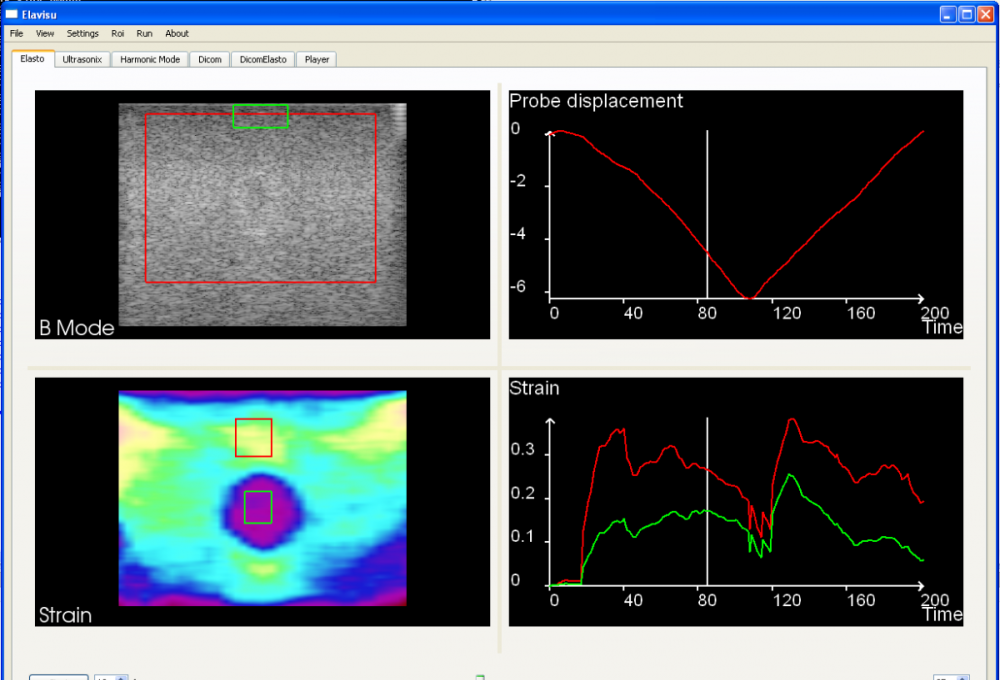
Elavisu's main window. Top left: B-Mode image (input). Top right: displacement of the probe. Bottom left: strain image and user-selected regions of interest. Bottom right: mean strain in the regions of interest.
Download:
Windows version. 1/Execute the installer. 2/Run Elavisu. 3/From the GUI select File Open demo file 'demo01.rf' in data_demo.
Using Elavisu on an ultrasound phantom.
Team
Project leader : Philippe Delachartre
Developers : Laurent Guigues, Tanguy Maltaverne, Andre Machado, Laurent Favreau, Eduardo Davila