Scientific and medical context :
The research lead for more than ten year about brain segmentation based on IRM data (T1 and T2 sequences) enable the measurement of cerebral and cerebelus structures volume. These variables are particularly interesting because they are correlated with the motor and cognitive development of the premature baby. These previous results are very encouraging, but unfortunately the access to IRM is limited.
Another way to image the neonate brain is cranial ultrasonography which can be realize through the anterior fontanel :
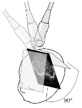
This medical exam can be realise directly at the patient bed and repeat as many times as necessary, moreover the access to sonography device is usually not a problem today. To the best of our knowledge, there is no way to automatically assess the volume of all the cerebral and cerebellar structures from sonography images.
The aim of this project is to develop new methods to segment the whole neonate brain using 3D ultrasound data. Such tools would provide quantitative measurements and temporal follow-up of the brain development which could help doctors to diagnose pathology.
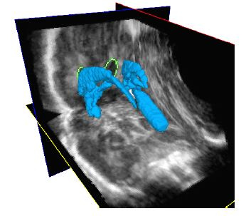
The quantification of lateral ventricles volume can improve the diagnosis and prognosis of brain anomalies. In the current clinical routine, a diagnosis is made based on 2D images of the brain, but the no quantitative measurement of the ventricules volume is made. To get this measurement, we developed the semi-automatic segmentation method describe below which need only 3 initial points :
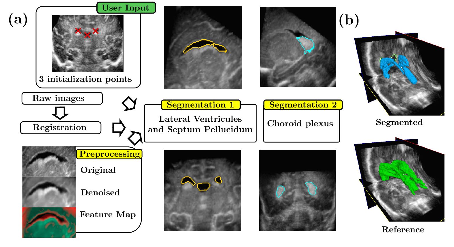
If you want to know more about this work, please read our IUS conference paper :
B. Sciolla, Martin, M., Quetin, P., et Delachartre, P., « Segmentation of the lateral ventricles in 3D ultrasound images of the brain in neonates », in IEEE International Ultrasonics Symposium, 2016.