- Participants
P. Clarysse, N. Duchateau, M. Sigovan (eq. 1), P. Delachartre (eq. 3)
- National and international collaborations
Wanyu LIU, METISLAB (Chine)
- Question
How to accurately quantify and analyze motion in dynamic cardiovascular image sequences? How to make possible comparison between individuals or longitudinal tracking of an individual ?
- Objective
To develop or adapt methods for the accurate quantification of motion and strains from dynamic image sequences, and for the registration of images and models to allow comparison between individuals or longitudinal tracking of an individual. Those methods are notably applied for the assessment and analysis of motion and deformation of anatomical structures in dynamic cardiovascular imaging.
- Method
- Motion quantification in image sequences. Our approaches are based on the concept of 2D analytical signal and the associated spatial phase information. A 2D sub-pixel displacement is deduced from phases integrated in local bilinear motion model in [WANG-15a]. In [Gao-16], a method, initially developed for motion tracking in color image sequences, has been adapted by integrating local phase, orientation and magnitude measurements that characterize a 2D monogenic signal. A more conventional differential approach that insures local intensity and shape features invariance to establish the correspondence obtained good results on both cardiac tagged-MR Imaging and Cine, the latter being part of the standard clinical protocol [Wang-19].
- Inter and intra-individual data registration. Two strategies are considered: if a parameterized segmentation is available for each subject/sample, data registration is based on a local coordinate system associated to an interpolation scheme [RUMI-17]. On the contrary, a registration between images or models is performed. The locally defined Cardiac function descriptors are mapped to the reference anatomy via this registration, using interpolation for scalar data, eventually completed by a reorientation for vector or tensor data [DUCH-11].
- Results and illustrations
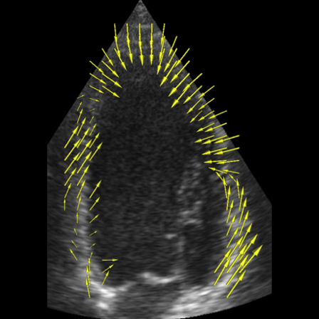
Figure 1 illustrates motion and radial strain quantification from cine cardiac MRI, used in clinical routine, and tagged MRI of the same patient. Both approaches shows a reduced contractile activity in IL, I and IS sectors where pathology has been identified, with slightly different distributions but with concordant sectorial values. Conventional clinical MRI sequences may therefore be used to assess regional cardiac function.
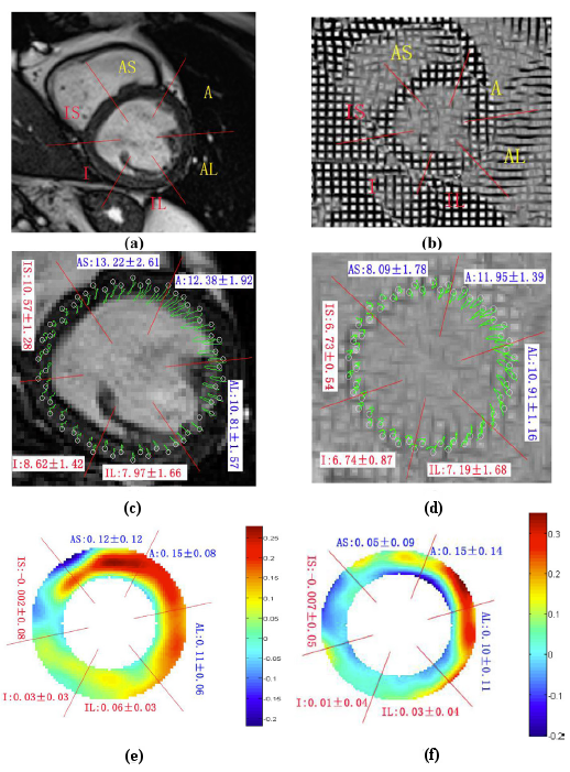
Figure 1. Motion and strain quantification in cardiac MRI. Short axis views in cine MRI (a) and tagged MRI (b) of the heart of an ischemic patient (end-diastole). (c-d) trajectories of points tracked over the cardiac cycle obtained with methods introduced in [Gao-16] for cine MRI (c) and in [WANG-15a] for tagged MRI. (e-f) cartographies of the radial deformation at end-systole. Mean and standard deviation per sector are indicated (identified pathological sectors are in red).
- Financial support
Works in this action are conducted within the framework of the associated international Laboratory METISLAB located in Harbin (China).
- Bibliographic References
- Journals
[Wang-19] L. Wang, P. Clarysse, Z. Liu, B. Gao, W. Liu, P. Croisille, and P. Delachartre, "A gradient-based optical-flow cardiac motion estimation method for cine and tagged MR images," Med Image Anal, vol. 57, pp. 136-148, 2019.
[Gao-16] B. Gao, Liu, W., Wang, L., Liu, Z., Croisille, P., Delachartre, P., et Clarysse, P., « Estimation of Cardiac Motion in Cine-MRI Sequences by Correlation Transform Optical Flow of Monogenic Features Distance », Physics in Medicine and Biology, vol. 61, p. 8640-8663, 2016.
[WANG-15a] L. Wang, Basarab, A., Girard, P. R., Croisille, P., Clarysse, P., et Delachartre, P., « Analytic signal phase-based myocardial motion estimation in tagged MRI sequences by a bilinear model and motion compensation », Medical Image Analysis, vol. 24, p. 149-162, 2015.
[DUCH-11] N. Duchateau, M. De Craene, G. Piella, E. Silva, A. Doltra, M. Sitges, et al., "A spatiotemporal statistical atlas of motion for the quantification of abnormal myocardial tissue velocities," Medical Image Analysis, vol. 15, pp. 316-328, 2011.
- Conferences
[RUMI-17] G. K. Rumindo, N. Duchateau, P. Croisille, J. Ohayon, and P. Clarysse, "Strain-Based Parameters for Infarct Localization: Evaluation via a Learning Algorithm on a Synthetic Database of Pathological Hearts," in Functional Imaging and Modeling of the Heart (FIMH), Toronto, Canada, 2017, p. accepted.
- PI (Intellectual property)
A French patent has been obtained on the approach based on the 2D analytical signal concept and a chinese one on a motion estimator based on fractional order differential:
[Dela-13]P. Delachartre, Basarab A., Clarysse, P., et Croisille, P., « Procédé d'estimation du mouvement dans une séquence d'images dynamiques », 2013.
[Liu-14] Liu Wanyu, Gao Bin, Kuai Zixiang, Clarysse Patrick « Movie nuclear magnetic resonance image sequence motion field estimation method based on fractional order differential », Cn Patent 103927725 - 16.07.2014.