- Participants
M. Orkisz, E.-J. Courtial, S. Qorchi (team 1), D. Vray, H. Liebgott, V. Perrot (team 3)
- National and international collaborations
A. Sérusclat, Ph. Moulin (HCL)
G. Zahnd (Technische Universität München , Munich, Allemagne)
A. van der Lugt (Erasmus MC, The Netherlands)
M. Skilton (Univ. Sydney, Australia)
- Question
Early detection of modifications in arterial-wall mechanical properties. Cardiovascular-risk stratification.
- Objective
Two-dimensional arterial-wall tissue-motion estimation during the cardiac cycle.
Estimation of compressions and shearing motion in the arterial wall.
- Methodology
Block matching, Kalman filtering, classification of curve shapes.
Detection of salient points, matching constrained by a model.
Adapted filtering, front propagation, coût minimum-cost path, dynamic programming.
- Results
- Motion estimation of the intima-media complex in the carotid artery
Progressive attenuation of the longitudinal-motion amplitude along the artery with a coefficient of -2.5 ± 2.0%/mm [bib]ZAHN-15a[/bib] (fig.1).
Te same motion-estimation method allowed experimental assessment of mechanical properties of vascular phantoms built using silicones [bib]COUR-15a[/bib].
Sensitivity and specificity of the order of 70% when classifying subjects as healthy or at risk based on carotid artery wall motion curve shapes, estimated from ultrasound image sequences [bib]QORC-17d[/bib].
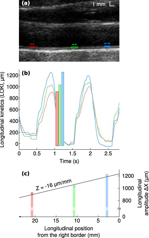
Figure 1. Example of longitudinal motion evaluated in a healthy subject at three different points along the carotid artery wall and showing a decrease in motion amplitude as the distance from the heart increases.
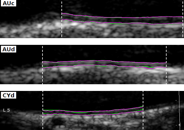
- Segmentation of the carotid-artery intima-media complex
With interactive initialization, segmentation errors in the first frame of each sequence respectively were of 29 ± 27 μm, 42 ± 38 μm, et 22 ± 16 μm for the lumen-intima and media-adventitia interfaces, and for the intima-media thickness (IMT). These uncertainties were of the same order as the inter- and intra-observer variabilities. The amplitude of the IMT temporal variations during the cardiac cycle was significantly greater in at-risk patients than in healthy volunteers (79 ± 36 vs. 64 ± 26 μm, p = 0.032) [bib]ZAHN-14[/bib].
With a fully automatic method, using the local thickness as additional dimension during the minimum-cost-path search, and validated on a larger and more diversified cohort, the respective segmentation errors were of 47 ± 70 μm, 55 ± 68 μm, and 66 ± 90 μm. The amplitude of the IMT temporal variations during the cardiac cycle also was significantly greater in at-risk patients than in healthy volunteers (106 ± 48 vs. 86 ± 34 μm, p = 0.001) [bib]ZAHN-17a[/bib].
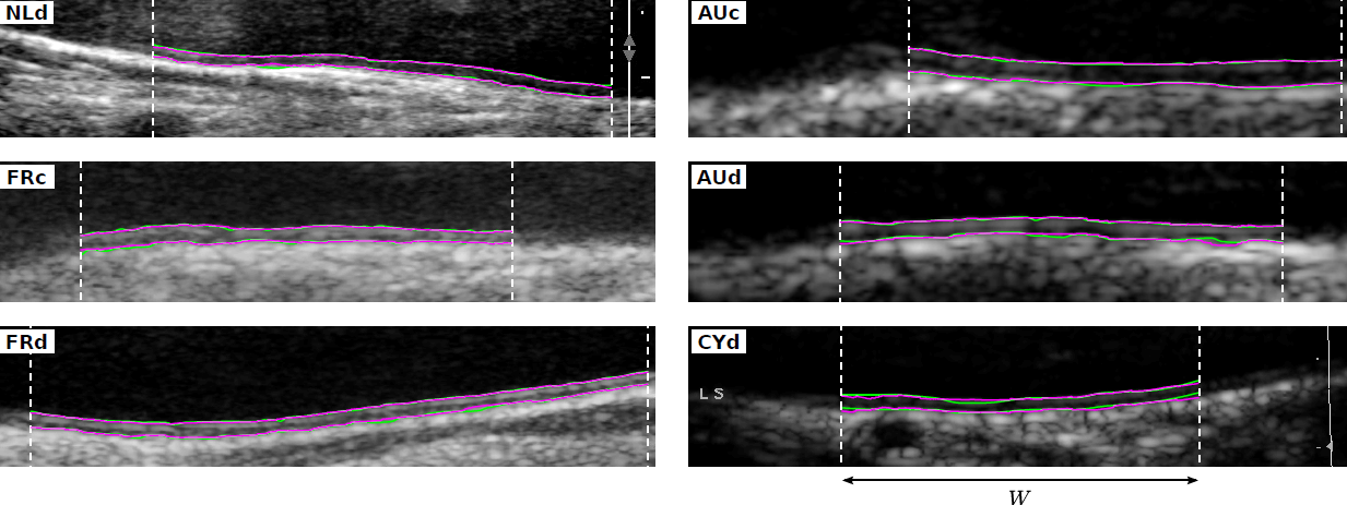
Figure 2. Results of the segmentation method (purple lines) compared to the reference contours traced by an expert (green lines), in subjects with varying image quality from six cohorts.
- Motion Estimation Refinement and Trajectory Classification
Motion estimation builds on block matching with a Kalman filter updating the reference-block gray levels, and incorporates a Kalman filter controlling the trajectory via a model using cosine decomposition. The estimated motion patterns were provided as input features to a machine-learning-based classifier that automatically assigned healthy or at-risk labels. Evaluated on 113 subjects, classification achieved 70% sensitivity and 72% specificity[bib]QORC-17d[/bib].
- Publications of the team
- Journals: [bib]ZAHN-17a[/bib], [bib]QORC-17d[/bib], [bib]COUR-16[/bib], [bib]COUR-15a[/bib], [bib]ZAHN-15[/bib], [bib]ZAHN-15a[/bib], [bib]ZAHN-14[/bib].
- Conferences: [bib]ZAHN-19[/bib], [bib]QORC-17b[/bib], [bib]QORC-17a[/bib], [bib]ZAHN-17b[/bib], [bib]COUR-15b[/bib], [bib]COUR-14a[/bib], [bib]COUR-14b[/bib], [bib]ZAHN-14a[/bib].
- IP (Intellectual Property)
CAROLAB Software (G. Zahnd) APP deposit: IDDN.FR.001.080024.000.S.P.2016.000.10000.