With the development of pulse wave imaging (PWI), the velocity of the so-called pulse wave (PWV) could be estimated, providing a quantitative evaluation of carotid stiffness. During the PhD work of Sébastien Salles, we developed original acquisition and image processing methods combining ultrafast US imaging, Transverse Oscillation tagging and Phase Based Motion Estimator algorithm.
These original methods have been implanted on our research systems to process RF images and validated on home-made phantoms, in collaboration with A. Yu, Hong-Kong University [SALL-15, IEEE Transactions on Ultrasonics, Ferroelectrics, and Frequency Control] and in collaboration with Lamcos Laboratory INSA, BQR funding [PERR-17b, IRBM]. We also studied volunteers and elderly patients [ZAHN-15, Medical Physics], as well in 3D [SALL-15b, IEEE Transactions on Ultrasonics, Ferroelectrics, and Frequency Control].
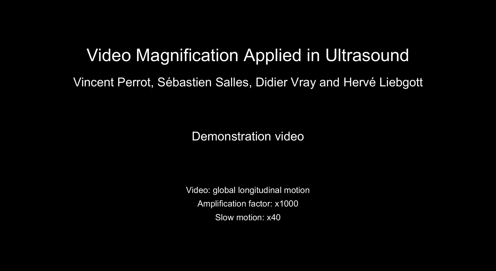
To enhance 2D subtle motion of the carotid wall which is not detected with a naked eye, we developed a visualization method able to see the Pulse Wave travel. Our Wave Magnification algorithm publication was selected as “highlights” by the journal editor [PERR-18a, IEEE Transactions on Biomedical Engineering].
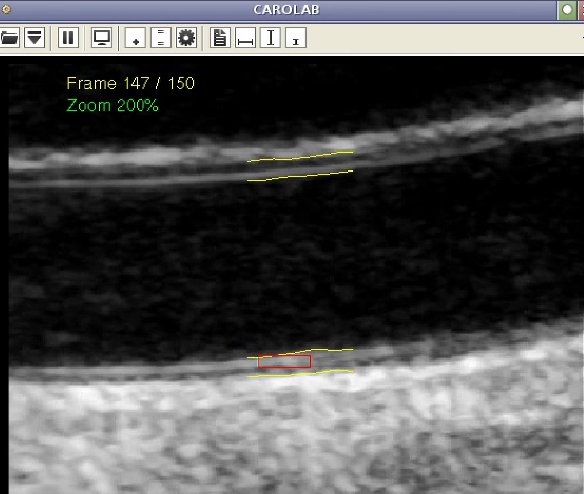
In parallel, in collaboration with team 2 (Maciej Orkisz) and the co-supervision of the PhD thesis of Guillaume Zahnd and Sami Qorchi, we developed B-mode sequence processing directly exported from clinical systems at the hospital. Carotid stiffness quantification was assessed by the tracking of salient speckle patterns of the carotid wall [ZAHN-15b, Ultrasound in Medicine & Biology], [ZAHN-14, Int J Comput Assist Radiol Surg], [CAROLAB software, https://www.creatis.insa-lyon.fr/carolab/], [QORC-17d, IRBM].