This work is done in the context of osteoarticular disease, and in particular for the understanding of bone fragility disease such as osteoporosis. To this aim, we are investigating new tomographic imaging techniques covering different scales. Capitalizing on our long term collaboration with the ESRF (the European Synchrotron, Grenoble), we have been addressing reconstruction problems in X-ray phase CT imaging and its applications for the investigation of bone tissue up to the cellular scale. In addition, we are working at the patient scale for the improvement of the characterization of bone micro-architecture in vivo, with the development of super resolution techniques. These works have been supported by the European Spatial Agency project ERISTO, the ANR project MULTIPS (2014-2018), the CLARA project Oncostarter Metangiobone (2016), a Long Term Project on beamline ID16A at the ESRF involving international partners (2014-2018), and 19 accepted beamtime proposals at ESRF.
Past actions :
The objective of this work is the quantification of the micro-architecture and quality of bone tissue by means of innovative imaging techniques. The ultimate aim of this research is to have a better understanding of the physiopathology of bone tissue in relation to its biomechanical properties and to improve the diagnosis or therapeutic follow-up of bone diseases.
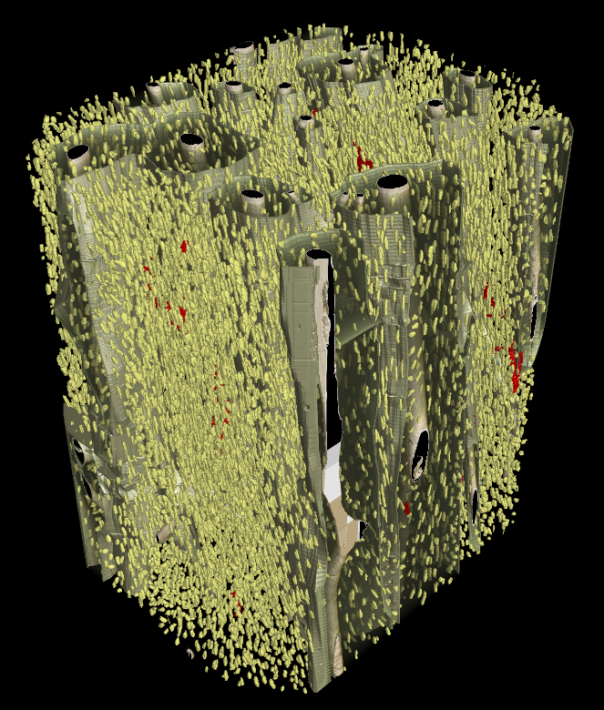
A large part of the research is done in collaboration with the ESRF (European Synchrotron Radiation Facility) in Grenoble, with which a very high resolution synchrotron radiation micro-CT system
Our research actions are largely based on this almost unique technique, which is used as a reference both for fundamental studies on bone tissue and for validation of emerging techniques for the diagnosis of osteoporosis. was developed (thesis by M. Salomé-Pateyron, 1998). The first images obtained on bone samples have opened up promising perspectives, both for the study of bone tissue and various materials.
Medical context
Osteoporosis is a bone fragility disease characterized by a low bone mass and alterations of bone micro-architecture leading to spontaneous bone fractures (vertebrae and femoral neck). It is estimated that 30 to 40% of persons over 60 years old will suffer from osteoporosis. Since the frequency of the disease is increasing with ageing, its management represents a major source of expense for public health.
The diagnosis of osteoporosis is currently mainly based on bone densitometry performed by dual x-ray absorptiometry and which measures bone mass. However, although a reduction in bone mass plays a major role in bone fragility, it is an insufficient predictor of the risk of fracture at the individual level.
In addition to bone quantity, other factors designated as “bone quality” are now recognized to play a role in the mechanical resistance to fracture. While bone quantity is a simple factor, bone quality, includes bone micro-architecture and many other parameters of the bone material which are still difficult to evaluate.
Our studies are based on three-dimensional imaging at different scales in order to assess bone quality.