Nous avons récemment développé et validé une méthode d’imagerie moléculaire de l’inflammation à l’aide de l’IRM haute résolution du petit animal. Cette approche repose sur le marquage des macrophages avec des nanoparticules superparamagnétiques injectées par voie intraveineuse. En regard des premiers résultats, il est possible de préciser la dynamique du processus inflammatoire post-ischémique sur un modèle murin. Cette étude expérimentale
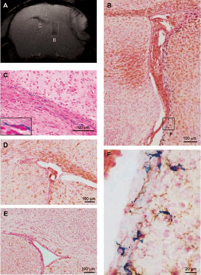
Figure. Correlation of gradient-echo (GRE) signals with immunohistology. A- GRE MRI 72h post-pMCAO and i.v. injection of USPIO. Note the hyposignal around the lesion, the contralateral corpus callosum and the ipsilateral peri-ventricular area. B- Double staining with Prussian blue and F4/80 of the right (ipsilateral) ventricle. Iron-stained microglia/macrophages are clearly visible along the lateral wall, in correlation with the hyposignal observed in GRE images (A). C- Prussian blue staining for iron in the contralateral corpus callosum of the corresponding slice. Positive staining was observed around cell nuclei, which suggests cytoplasm uptake (insert). D- F4/80 immunostaining for mouse microglia/macrophages in the contralateral corpus callosum. Note the F4/80+ brown cells, in spatial agreement with iron+ cells (B) and MRI hyposignal (A). E- F4/80 immunostaining of a non-operated control mouse in the left corpus callosum, with no positive staining. F- Double staining with Prussian blue and F4/80: magnification of B. Microglia/macrophages as identified by their brown color and typical ramified shape, were also blue-stained, suggesting USPIO intra-cellularity.