COORDINATEUR SCIENTIFIQUE: Pierre Croisille, PUPH, Chef de Pole IMOFON (CHU de Saint-Etienne), Chef de departement (Radiologie et Medecine Nucléaire), Chef de Service de Radiologie au CHUSE
Membres du Projet: Equipe MYRIAD: P. Clarysse, N. Duchateau, O. Bernard, T. Grenier, M. Sdika. Equipe ULTIM: H. liebgott, F. Varray. Equipe MAGICS: O. Beuf, P. Croisille, L. Petrusca, C. Bouton, W. Romero, E. Van Reeth, K. Tse Ve Koon, B. Leporq, H. Ratiney, M. Viallon.
I. Objectifs du projet
Ce projet vise à la réalisation des développements technologiques et méthodologiques pour permettre l'imagerie multimodale multiparamétrique quantitative du cœur et du muscle squelettique (PET / MR, mais aussi des US / MR), y compris de nouvelles stratégies théranostiques dans un environnement clinique. Ce projet permettra de répondre à ces défits par une approche multidisciplinaire impliquant des physiologistes, des physiciens des US et de la RMN, des informaticiens des laboratoires régionaux, ainsi que des partenaires français et industriels (SIEMENS, OLEA, ...), tout en renforçant les collaborations nationales et internationales existantes (Départements de Radiologie, cardiologie, laboratoires de recherche (CRMBM-CEMEREM, UMR CNRS 7339 (Marseille), les experts en imagerie cardiaque des Universités européennes (Lausanne Suisse), allemande (Bremen) et américaines (Los Andes(Colombia), UCLA( Los Angeles,California), UVA (Virginia), Harvard (Boston, MS)...)).
II. Contexte socio-économique
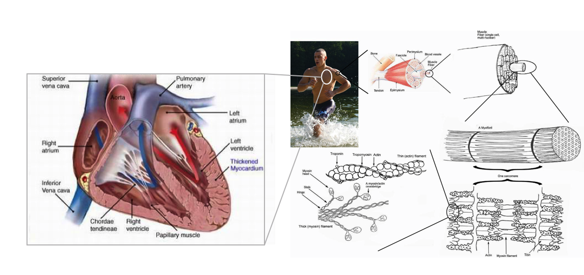
Muscle cardiaque: Parmi les maladies du muscle cardiaque, les cardiopathies ischémiques restent la première cause de mortalité dans le monde, avec une progression de 21 % au cours de la dernière décennie (Roth et al, lancet 2018). En France, la mortalité à 10 ans après un infarctus du myocarde (IM) est tombée à 10 % grâce à des efforts multiformes, notamment des réseaux régionaux pour une intervention coronarienne plus rapide, des soins intensifs spécialisés après un IM, des efforts itératifs d'éducation de la population pour réduire les facteurs de risque et reconnaître les symptômes précoces (initiatives conjointes des agences nationales françaises de santé publique et des sociétés de cardiologie au cours des deux dernières décennies) (Grave C et al. 2019). Pourtant, les maladies cardiovasculaires (MCV) restent un problème majeur de santé publique. Avec un plus grand nombre de survivants après un IAM, environ 60 % des cas d'insuffisance cardiaque (IC) en France ont une étiologie ischémique, et le coût associé à la prise en charge de l'IC ischémique est estimé à 3,6 milliards d'euros par an en France. L'insuffisance cardiaque devrait augmenter de 25 % tous les quatre ans dans les années à venir en raison du vieillissement de la population.
- Santé Publique France. Infarctus du myocarde. 2019. https://www.santepubliquefrance.fr/maladies-et-traumatismes/maladies- cardiovasculaires-et-accident-vasculaire-cerebral/infarctus-du-myocarde/le-scan/#tabs.
2. Société Française de Cardiologie /Groupe Insuffisance Cardiaque (GICC). Livre Blanc: Pladoyer pour la prise en charge de l’insuffisance cardiaque et des cardiomyopathies. 2021. https://www.chu-clermontferrand.fr/sites/default/files/media/2021- 11/Livre-blanc-de-lInsuffisance-Cardiaque.pdf.
Le Muscle squelettique: Le muscle squelettique (c'est à dire nos muscles des cuisses, dos, jambes, bras et...) est l'organe et allié majeur de notre santé. Au-delà de la mobilité, son activité a une action protectrice, voire curative, sur l’organisme, d’où l’importance de le mobiliser à tout âge, même lorsque l’on est concerné par une maladie. Mais qu’il soit sain, malade ou entraîné, il est loin d’avoir révélé tous ses secrets. Le muscle et ses bienfaits sont encore trop peu reconnus, pourtant cet organe est un allié majeur de notre santé. Au-delà de la mobilité, son activité a une action protectrice, voire curative, sur l’organisme, d’où l’importance de le mobiliser à tout âge, même lorsque l’on est concerné par une maladie. Mais qu’il soit sain, malade ou entraîné, il est loin d’avoir révélé tous ses secrets.
Les recherches doivent se poursuivre et notemment en imagerie, pour comprendre chacun de ses mécanismes, une nécessité en matière de santé publique, très bien mise en lumière, le 1er juin 2023, lors des Assises du muscle qui ont ouvert la première Semaine du muscle, organisées par l’Institut de Myologie et l’AFM-Téléthon.
1. https://www.afm-telethon.fr/sites/default/files/media/documents/AFM_VLM_206_BD_DOSSIER.pdf
III. Les moyens du projets
Le projet a obtenu un financement FEDER, Conseil Général 42 (CG42) et Saint-Etienne Métropole (SME) pour se doter d'un générateur de puissance 512 Voies VERASONIC. Le projet dispose d'un puissante Plateforme de recherche clinique combinant un IRM 3T équipée d'un chaine multi-noyau et d'un ergomètre MR compatible doté des module cardiaque et quadriceps. En savoir plus...
Lien avec les Equipements et Investissements d'avenir: Equipex LILI (plateforme PET-MR plateforme), LabEX Primes, LabEX Celya.
Financement obtenus: Financement MESSIDOR (IGRESP INSERM 2025), France Life Imaging (FLI) WP4 (SEISM, COLCAR), FLI WP3(TSONOAMI), Dotation de Recherche René Foudon, Fédération française de cardiologie (FFC, 2018) RHU MARVELOUS (2016), PHRC MARVEL (2016). Fond européen FEDER (2014), CG42, SME. Co-financements: CARIM, DIAPASON.
Partenaires industriels: OLEA Medical, Siemens, Circle, Carestream, Supersonic Imagine.
Programme et présentations powerpoints des workshops accessibles aux membres actifs du Projet sur l'intranet de CREATIS au lien suivant: En savoir plus...
V. Les Sous-Projets en cours
MUST: Le projet MUST a pour objectif d'aider à comprendre l'effet de l'ultra-endurance sur l'organisme et son impact au niveau musculaire et cardiaque. L'équipe (P. Croisille (PI, Team #5), O.Beuf (Team #5), M. Viallon (Team #5), H. Ratiney (Team#5) B. Leporq (Team#5) T. Grenier (Team#2)) a choisi de mener cette étude lors de l'Ultra-Marathon de Montagne le plus extrême au monde, Le Tor des Géants: 330 km, 24.000 m de dénivelé positif d'une seule traite (soit 3x l'Everest), dans la Vallée d'Aoste, en Italie et au pied du Mont-Blanc. Cette épreuve de tous les superlatifs a eu lieu du 7 au 14 Septembre 2014.
TSONOAMI (Imagerie Thérapeutique et infarctus du myocarde à la phase aigüe): Développements Méthodologiques US-MR. M. Viallon (PI,Team #5), P. Croisille (Team #5), L. Petrusca (Team#5), O.Beuf (Team #5), H. Liebgott (Team#3). Il existe un attrait clinique élevé pour les nouvelles technologies théranostiques, non-invasives et capable d’offrir une efficacité au moins comparable à celui des traitements conventionnels (produits thérapeutiques et diagnostiques). Ultrasons focalisés (FUS) a déjà un large impact dans le domaine médical, les différentes applications cliniques étant identifiés à ce jour pour les différents organes (sein, de la prostate, les os, la fibrose bénigne de l'utérus). En raison des défis importants, les applications de FUS pour les thérapies cardiaques n'a pas encore été étudié. Il y a un intérêt majeur pour étudier les mécanismes et dans quelle mesure les ultrasons pourraient atténuer la réponse inflammatoire dy myocarde et y remédiée. Les effets bénéfiques et donc soi-disant effet «sono-cardioprotecteur" des US exploreront le drainage mécanique de massage par une force de rayonnement acoustique (ARF) ou sur l'amélioration de la microcirculation mycoardium ischémique. Il y a aussi un intérêt évident dans le contexte des thérapies guidées par l'image dans les organes mobiles, pour développer l'imagerie hybride US-MR.
PET-MR Quantitative Perfusion, Team 5 - P. Croisille (PI, Team #5), O.Beuf (Team #5), M. Viallon (Team #5), C. Daviller(Team#5), C. Frindel (Team #2), T. Grenier (Team #2),. (Collaboration: RHU MARVELOUS (OLEA Medical, CARMEN U1060, Cynbiose), Pr Marc Janier(HCL), SIemens). Establishing normal and pathology related Myocardial perfusion standard for improved patient profiling based on newly established Bio-Atlas provided by Advanced Imaging Techniques and Biomarkers. The objective of this collaboration is 1) acquire the methodology to achieve quantitative values of Myocardial Blood Flow using MRI 2) to utilize advanced imaging methods (simultaneous PET-MRI) to investigate absolute perfusion values in humans, and relate them to the cardiovascular metabolic state. Our ultimate goal is to establish a predictive bio-atlas based on advanced imaging biomarkers, and to make available gold standard reference for clinical risk factors investigation, prediction, and improved objective patient management.
Biomechanics of the Heart: A Model-Based Non-Invasive Regional Identification of Myocardial Diseases : P. Clarysse (PI, Team #1), N. Duchateau (Team #1), P. Croisille (Team #5), M. Viallon (Team #5), O. Bernard (Team #2), C. Lartizien (Team #2), P. Delachartre (Team #3) in Collaboration with TIMC-Grenoble, ICJ-Lyon, CARMEN-Lyon. The aim of this sub-project is to introduce and validate a new strain based model and methods for the non-invasive regional characterization of the myocardial viability. New concept of myocardial fiber tonicity (MFT) is obtained from the instanciation of a biomechanical parametric finite element (FE) model of the left ventricle (LV), including fiber architecture to patient strain and pressure measurements. The methodology is general, but will be first experimented in cardiac MRI and US, and should provide new biomarkers of regional estimation of the amount of active tension within the myocardium, that may reflect the effective health status of the tissue. Strain measurements will be obtained with state of the art dynamical tagged-MRI (Collab. Prof. Stuber, EPFL).
VI. Les Publications du Projet IDM4
1: Automatic myocardial ischemic lesion detection on magnetic resonance perfusion weighted imaging prior perfusion quantification: A pre-modeling strategy. Daviller C, Grenier T, Ratiney H, Sdika M, Croisille P, Viallon M. Comput Biol Med. 2019 Jul;110:108-119. doi: 10.1016/j.compbiomed.2019.05.001. Epub 2019 May 2. PubMed PMID: 31153004.
7. Shear-Wave Elastography Assessments of Quadriceps Stiffness Changes prior to, during and after Prolonged Exercise: A Longitudinal Study during an Extreme Mountain Ultra-Marathon. Andonian P, Viallon M, Le Goff C, de Bourguignon C, Tourel C, Morel J, Giardini G, Gergelé L, Millet GP, Croisille P. PLoS One. 2016 Aug 31;11(8):e0161855. doi: 10.1371/journal.pone.0161855. PMID: 27579699
8. Quantifying the effect of tissue deformation on diffusion-weighted MRI: a mathematical model and an efficient simulation framework applied to cardiac diffusion imaging. Mekkaoui I, Moulin K, Croisille P, Pousin J, Viallon M. Phys Med Biol. 2016 Aug 7;61(15):5662-86. doi: 10.1088/0031-9155/61/15/5662. PMID:27385441
9. Comparison of Immediate With Delayed Stenting Using the Minimalist Immediate Mechanical Intervention Approach in Acute ST-Segment-Elevation Myocardial Infarction: The MIMI Study. Belle L, Motreff P, Mangin L, Rangé G, Marcaggi X, Marie A, Ferrier N, Dubreuil O, Zemour G, Souteyrand G, Caussin C, Amabile N, Isaaz K, Dauphin R, Koning R, Robin C, Faurie B, Bonello L, Champin S, Delhaye C, Cuilleret F, Mewton N, Genty C, Viallon M, Bosson JL, Croisille P; MIMI Investigators*.Circ Cardiovasc Interv. 2016 Mar;9(3):e003388. doi: 10.1161/CIRCINTERVENTIONS.115.003388. PMID:26957418
10. Image-Based Investigation of Human in Vivo Myofibre Strain. Wang VY, Casta C, Zhu YM, Cowan BR, Croisille P, Young AA, Clarysse P, Nash MP. IEEE Trans Med Imaging. 2016 Nov;35(11):2486-2496. PMID: 27323360
11. Myocardial Extracellular Volume Estimation by CMR Predicts Functional Recovery Following Acute MI. Kidambi A, Motwani M, Uddin A, Ripley DP, McDiarmid AK, Swoboda PP, Broadbent DA, Musa TA, Erhayiem B, Leader J, Croisille P, Clarysse P, Greenwood JP, Plein S. JACC Cardiovasc Imaging. 2016 Oct 18. pii: S1936-878X(16)30645-3. doi: 10.1016/j.jcmg.2016.06.015. [Epub ahead of print] PMID:27771398. Free Article
12. In vivo free-breathing DTI and IVIM of the whole human heart using a real-time slice-followed SE-EPI navigator-based sequence: A reproducibility study in healthy volunteers. Moulin K, Croisille P, Feiweier T, Delattre BM, Wei H, Robert B, Beuf O, Viallon M. Magn Reson Med. 2016 Jul;76(1):70-82. doi: 10.1002/mrm.25852. PMID:26301785
13. Effects of glycaemic variability on cardiac remodelling after reperfused myocardial infarction: Evaluation of streptozotocin-induced diabetic Wistar rats using cardiac magnetic resonance imaging. Joubert M, Hardouin J, Legallois D, Blanchart K, Elie N, Nowoczyn M, Croisille P, Coulbault L, Bor-Angelier C, Allouche S, Manrique A. Diabetes Metab. 2016 Mar 10. pii: S1262-3636(16)30012-X. doi: 10.1016/j.diabet.2016.02.002. [Epub ahead of print]. PMID:26971835
14. A new look at left ventricular remodeling definition by cardiac imaging. Bière L, Donal E, Jacquier A, Croisille P, Genée O, Christiaens L, Prunier F, Gueret P, Boyer L, Furber A. Int J Cardiol. 2016 Apr 15;209:17-9. doi: 10.1016/j.ijcard.2016.02.009. No abstract available. PMID:26878466