Proton imaging has been a major field of research of the past five years, funded by the Labex PRIMES, a ministry PhD grant, a Marie Curie postdoc fellowship, the Fondation pour la Recherche Médicale and a franco-bavarian project for the collaboration with the Ludwig Maximilian University (LMU, Munich). The difficulty in proton imaging is that each proton follows a different path due to multiple Coulomb scattering (MCS) which can be corrected for using a system recording the position and direction of each proton, both before and after the object. Nicolas Arbor (postdoc) has demonstrated the interest of this modality for proton therapy [Arbor-2015- Physics in Medicine and Biology]. In her PhD thesis, Catherine Quiñones has investigated the use of an estimate of the most likely path (MLP) of each proton for alternative modalities of proton CT enabled by list-mode systems (attenuation and scattering) [Quinones-2016- Physics in Medicine and Biology] (proffered paper). Nils Krah’s postdoc has focused on more fundamental questions: achievable spatial resolution for various imaging set-ups [Krah-2018- Physics in Medicine and Biology] and regularization of an inverse problem fitting a proton radiography to an X-ray CT [Krah-2019- Physics in Medicine and Biology]. The on-going PhD work of Feriel Khellaf has demonstrated the accuracy of the MLP in heterogeneous media despite the homogeneity assumption using Monte Carlo simulations [Khellaf-2019- Physics in Medicine and Biology]. We have applied our investigations to integrated systems [Krah-2018- Physics in Medicine and Biology] and, in collaboration with LMU and Loma Linda (USA), list-mode systems [Dedes-2017- Physics in Medicine and Biology][Dedes-2018- Med Phys].
Proton imaging
The use of protons for medical imaging as an alternative to X-rays is not new but its interest has been renewed with the development of proton therapy since proton sources are more available and it can improve the precision of the treatment. In France, two centers treat patients using protons, in Orsay and Nice, and a new center has been built in Caen. A fundamental difficulty in proton imaging is multiple Coulomb scattering which causes the charged protons to follow a random curved path. The loss in spatial resolution can be circumvented by measuring the position and the direction of each proton, both before and after the patient, and by deducing its most likely path. We are developing new software methods to improve the quality of proton radiographies and computed tomographies (CTs), and to propose new treatment protocols based on these images.
We have developed a new filtered backprojection that can account for each proton most likely path, and we have demonstrated improved image quality for energy-loss and attenuation proton CT. Comparison with iterative reconstruction algorithms has shown similar spatial resolution. The benefit for proton therapy compared to the current clinical practice based on x-ray CT has been demonstrated using GATE Monte Carlo simulations (see figure below).
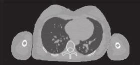
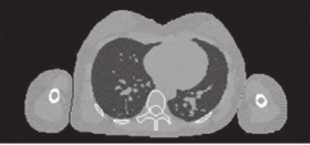
Simulated proton CT (left) and x-ray CT (right) of the ICRP computational female phantom. The proton CT image has been reconstructed using filtered backprojection reconstruction with estimates of the most likely path of each proton.
Recently, we have developed a framework to assess the spatial resolution of each possible proton imaging system. We are currently developing strategies that combine a proton radiography with in-room cone-beam CT to minimize proton imaging time. Our developments have been tested on real data of the phase II pCT prototype of Loma Linda (USA) for proton-by-proton data and a commercial range telescope in Orsay for integrated data. Our strong visibility in this niche field has led us to organize the first proton imaging workshop (https://protonimaging.sciencesconf.org/).
Software development
Most of the simulation works have been included in the open-source GATE managed by the international collaboration Opengate (http://www.opengatecollaboration.org).
- GATE: Medical physics Monte-Carlo simulation. Open-source. ~1000 users. [Sarrut et al. Med Phys 2014, Jan et al PMB 2011]. International collaboration. http://www.opengatecollaboration.org
- VV: 4D Medical image viewer. Open-source. ~100 users. [Rit et al 2011]. https://www.creatis.insa-lyon.fr/rio/vv