Elisabeth BRUSSEAU
 |
Position
: CNRS
Researcher (CNRS is the French National Center for Scientific Research)
Laboratory :
CREATIS UMR 5220, INSERM U1044
Mailing address :CREATIS
Bâtiment Blaise Pascal 7, avenue Jean
Capelle
69 621 Villeurbanne
FRANCE
Phone: +33
4 72 43 61 41
Email : elisabeth.brusseau@creatis.insa-lyon.frFax : +33 4 72 43 85 26 |
1. Research activities
Keywords : medical
imaging, signal and image processing, elastography, segmentation,
ultrasound.
2.
Ultrasound elastography
Ultrasound
elastography is an imaging technique dedicated to the
investigation of the mechanical properties of soft biological tissues.
This technique is of fundamental interest for the clinical diagnosis of
various diseases, since the development of a pathological process is
often correlated with local changes in tissue stiffness.
Elastography might therefore provide useful information for early
detection and characterization of various pathologies and for patient
treatment follow-up. For the technique termed "quasi-static
elastography", the tissue is generally deformed
by compressing it with the ultrasound probe. Tissue internal
displacements
and deformations are then locally estimated from acquired
radiofrequency (RF) ultrasound (US) images, by partitioning the US data
into many overlapping regions of interest (ROI) and by evaluating,
for each ROI, the positional variations induced by the stress.
Strain maps reveal tissue elasticity by providing information on tissue deformation under compression.
My main research works in quasi-static elastography focused on successively developing 1D, 2D and 3D strain estimation techniques, to image tissue deformation under load. We currently assess our technique in the clinical conditions with patients suffering from breast lesions, and with data acquired at the Hôpital de La Croix-Rousse, Hospices Civils de Lyon, Lyon, France or provided by the Institute of Cancer Research, Sutton, United Kingdom.
Results obtained with a 2D locally regularized strain estimation method [Brusseau et al., IEEE TMI 2008] during freehand examination are provided below.
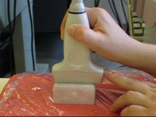 |
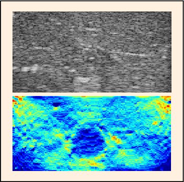
This
movie illustrates the way an elastographic experiment is lead during
freehand examination. 1- The medium under investigation is cautiously compressed with the ultrasound probe. 2- On the scanner screen, the medium deformation is observable. The dark inclusion of circular shape is clearly visible. However during the compression, ultrasound B-mode images exhibit similar patterns and brightness. A lesion undetectable in one ultrasound image may thus remain unvisible during the compression. 3- Strain images are computed from the acquired ultrasound data sets. Some examples are provided below. [Click on the picture to run the movie.] |
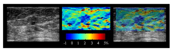 |
Case of a fibroadenoma (confirmed by hystology)
Left : conventional ultrasound image. Middle : Strain image. Right : Superimposition of the two images. |
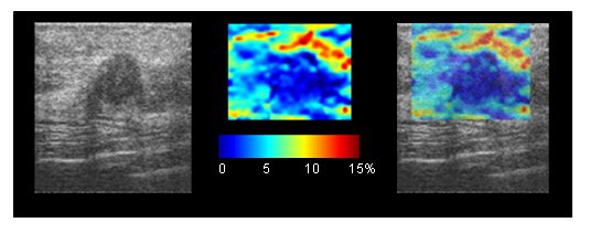 |
Case of a suspected carcinoma Left : conventional ultrasound image. Middle : Strain image. Right : Superimposition of the two images. These initial results are published in SPIE Medical Imaging 2011. |