Modelling skin surface areas involved in water transfer in the Palmate Newt (Lissotriton helveticus)
Abstract
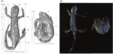
Magnetic resonance imaging (MRI) based 3D reconstructions were used to derive accurate quantitative data on body volume and functional skin surface areas involved in water transfer in the Palmate Newt (Lissotriton helveticus (Razoumovsky, 1789)). Body surface area can be functionally divided into evaporative surface area that interacts with the atmosphere and controls the transepidermal evaporative water loss (TEWL); ventral surface area in contact with the substratum that controls transepidermal water absorption (TWA); and skin surface area in contact with other skin surfaces when amphibians adopt water-conserving postures. We generated 3D geometries of the newts via volume-rendering by a “segmentation” process carried out using a graph-cuts algorithm and a Web-based interface. The geometries reproduced the two postures adopted by the newts, i.e., an I-shaped posture characterized by a straight body without tail coiling and an S-shaped posture where the body is huddled up with the tail coiling along it. As a guide to the quality of the surface area estimations, we compared measurements of TEWL rates between living newts and their agar replicas (reproducing their two postures) at 20°C and 60% relative humidity. Whereas the newts did not show any physiological adaptations to restrain evaporation, they expressed an efficient S-shaped posture with a resulting water economy of 22.9%, which is very close to the 23.6% reduction in evaporative surface area measured using 3D analysis.