(C. Revol-Muller)
Objectives
The goal of this work was to develop an automated method for the segmentation of three-dimensional IRM images. For most of segmentation methods, setting initial parameters is a difficult problem which directly governs the quality of the segmentation. When this setting is manual, results depends on user choices and can not be used for clinical study. The proposed method is a solution to this problem.
Methods
This method is based on a 3D region growing algorithm which differs from other techniques by reconsidering the merge of pixels into the region growing at each step. Initial seeds location is determined by the original image histogram and homogeneity criterion which governs the region growing is deduced from an evaluation function
Results
|
a) Slice from a bone MRI image (128x128x128) |
b) Slice segmented by our method |
c) Slice obtained using an automated binary threshold |
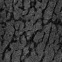
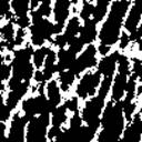
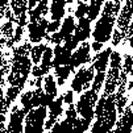
Fig.1: Comparison between our method (b) and an automated thresholding method (c).
|
a) |
b) |
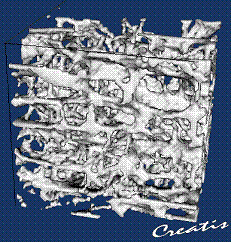
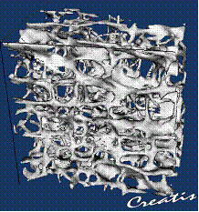
Fig.2 : Comparison between the resulting segmentation of our method applied to a MRI image (a) with the reference image provided by synchrotron tomography (ESRF) (b)
Two acquisitions of the same human calcaneum bone sample were made: by high resolution IRM and by synchrotron radiation (ESRF). MRI produces images with a resolution of 80 microns whereas synchrotron provides a resolution of 10 microns with a very high signal to noise rate. The ESRF image was under-sampled in order to work at the same spatial resolution as the MRI image.
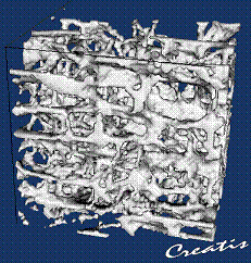
Fig.3 : Illustration of our segmentation applied to a MRI image