Overview
NEWS
13th of May 2024
Opening of the challenge: training / testing datasets and codalab online plateform are available
26th of July 2024
End of the challenge
Goal of the MYOSAIQ challenge
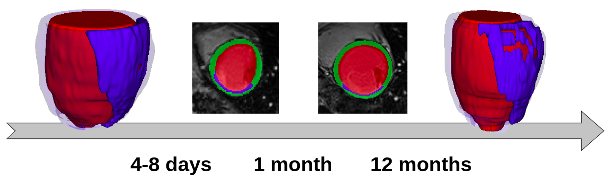
Late gadolinium enhanced magnetic resonance images (LGE) are used in clinical practice to quantify the areas of myocardial muscle affected following a myocardial infarction (MI). In this context, the aim of this challenge is twofold:
Originality of the MYOSAIQ challenge
The originality of this challenge lies in the analysis of a large database composed of 439 MR-LGE exams and integrating the main various issues encountered in daily clinical cardiac imaging, namely:
Late gadolinium enhanced magnetic resonance images (LGE) are used in clinical practice to quantify the areas of myocardial muscle affected following a myocardial infarction (MI). In this context, the aim of this challenge is twofold:
- compare the performance of automatic methods on the segmentation of the left ventricular endocardium and epicardium on LGE images;
- compare the performance of automatic methods for the quantification of MI lesion size, at different phases of the longitudinal evolution of the disease on images acquired at the acute phase (4-8 days post-MI & reperfusion therapy) and delayed phase (1 month / 12 months post-MI & reperfusion).
Originality of the MYOSAIQ challenge
The originality of this challenge lies in the analysis of a large database composed of 439 MR-LGE exams and integrating the main various issues encountered in daily clinical cardiac imaging, namely:
- the longitudinal evolution of the patho-physiology of the heart;
- the presence of image artifacts;
- the use of different MR acquisition techniques (3D versus 2D PSIR);
- the use of different MR acquisition settings from different centers.