Simone Balocco1,2, Olivier Basset1, Jacques Azencot1, Piero Tortoli2, Christian Cachard1
1 CREATIS, Université de Lyon , INSA de Lyon, Université Lyon 1, CNRS UMR 5220, INSERM U630, Lyon, France
2 Microelectronic Systems Design Laboratory, Università di Firenze, Italy
Aim of the study
To propose a 3D model reproducing the biomechanical behaviour of human blood vessels
Model
The model, based on a multilayer geometry composed of right generalized cylinders (RGCs), enables the representation of different vessel structures such as stenoses, bifurcations and the associated pathologies.
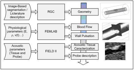
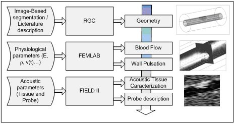
Flow chart illustrating the interaction between different modules (RGC, FEMLAB, FIELD II), of the 3D model.
The model is designed as a multilayer structure to control the vessel geometry as well as the intima–media thickness. Each area and each layer of the phantom can be described by independent sets of mechanical and acoustic properties and geometrical shapes. Different vessel structures, including bifurcations, with associated pathologies such as local arterial wall thickening, aneurysms, stenosis and atherosclerotic plaques can be simulated.
Method
Using a finite element approach, blood flow is simulated by considering a dynamic displacement of the scatterers, while arterial pulsation due to the hydraulic pressure is taken into account through a fluid–structure interaction based on a wall model. Each region is acoustically characterized using FIELD II software, which produces the radiofrequency (RF) echo-signals corresponding to echographic scans.
Applications and Results
Acoustic physiological phantoms of carotid arteries surrounded by elastic tissue are presented hereafter to illustrate the model’s capability.
The first corresponds to a healthy blood vessel, the second includes a 50% stenosis and the third represents a carotid bifurcation.
(a)(b) (c)
3D representation of blood vessel geometries obtained from RGC models. Phantoms represent a healthy vessel, (a), a stenosed vessel, (b) and a bifurcation branching vessel, (c). In (b) and (c) the external RGC is transparent to show the external artery layers.
(a)
(b)
(c)
Simulated B-mode of the healthy () (a), the stenosed () (b) and the bifurcating () vessel extracted from a sequence of images, frozen during the cardiac cycle in diastole for (a) and (b), and (c))
M-mode, B-mode and color Doppler images derived from these phantoms can be provided.
An example of identification of the atherosclerotic plaque boundaries on Doppler color images is reported.
Simulated Doppler at 10-kHz PRF (a) and corresponding reference velocity (finite element) images (b) showing blood recirculation downstream in the stenosed area. The simulated fluid flows from left to right. The geometry of the structure is superimposed on the Doppler image on (a).
The model can be used as a tool for the preliminary evaluation of ultrasound signal processing and visualization techniques [Balocco].