- Participants
I. Mirea (PhD Student), F. Varray, Y.-M. Zhu, L. Fanton, I.E. Magnin
- National and international collaborations
Creatis, teams 1,3 et 4 (F. Peyrin, M. Langer)
Metislab, Harbin (W.Y. Liu, Y.L. Zhang, S.L. Wang (PhD Student)
- Unresolved question
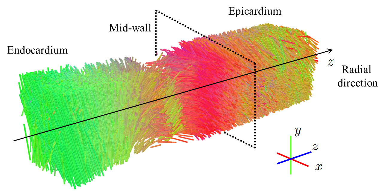
What is the 3D spatial organisation of the different components of human heart tissue at the cellular and tissue levels?
- Objective
Access to the microarchitecture of the components of the heart tissue of the cell (micrometer) scale to that of the body (centimeter).
- Method
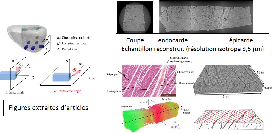
- Extract samples of fresh human heart wall (Left Ventricle)
- Reconstruct the samples using X-ray phase-contrast micro-tomography (2500 X-ray projections acquired at the Synchrotron at a micrometric resolution)
- Process the reconstructed volumes (multiscale segmentation)
- Analyse the binarised reconstructed volumes to quantify the features of the 3D microstructure and get the arrangement of the tissue components.
- Define new bio-marquers.
- Results and illustrations
- Grants
METISLAB (LIA CNRS-Inserm); Labex PRIMES ; INSA Lyon ; CREATIS ; HIT Harbin
- Publications
F. Varray, Mirea, I., Langer, M., Peyrin, F., Fanton, L., and Magnin, I. E., « Extraction of the 3D Local Orientation of Myocytes in Human Cardiac Tissue Using X-ray Phase-Contrast Micro-Tomography and Multi-Scale Analysis », Medical Image Analysis (38) p. 117-132, 2017, (IF = 4, 56).
I.Mirea, L.Wang, F. Varray, YM Zhu, E.E Davila-Serrano, I.E. Magnin, “Statistical Analysis of Transmural Laminar Microarchitecture of the Human Left Ventricle”, ICSP, Chengdu, 2016, pp 53-56.
I. Mirea, F. Varray, Y. M. Zhu, L. Fanton, M. Langer, P. Jouk, G. Michalowicz, Y. Usson, and I. E. Magnin "Very High-Resolution Imaging of Post-Mortem Human Cardiac Tissue using X-ray Phase Contrast Tomography", FIMH'15, Maastricht, LNCS 9126, pp. 172-179, 2015.
F. Varray, L. H. Wang, L. Fanton, Y. M. Zhu, and I. E. Magnin, "High resolution extraction of local human cardiac fibre orientations", FIMH’13, London, LNCS 7945, pp. 150-157, 2013.
- Protection
The data acquired at the ESRF will be available to the scientific community after 3 years.