
Purpose and context
Recently, the osteocyte system has received a lot of attention in the research of mechanisms governing bone fragility. Osteocytes, the most abundant bone cells, communicating through an extensive network of processes, are supposed to play a major role in bone mechanotransduction. They are encapsulated within a pore network, called the lacunar-canalicular network (LCN). Little quantitative data are found on the organization of the LCN due to the lack of methods to assess this structure. Imaging the LCN is challenging, due to its deep location within hard bone tissue, the small size of lacunae (a few micrometers) and canaliculi (100-700 nm in diameter) and the complexity of this network.
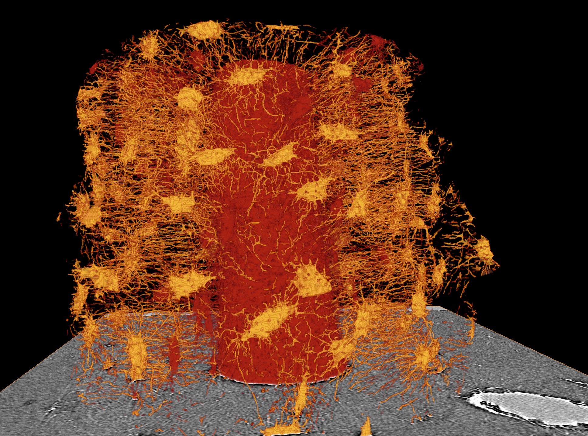
Figure 1 : Illustration of a the lacuna-canalicular network (yellow) around a whole osteon (red) obtained from SR micro-CT (voxel size 300 nm)
3D Micro-CT imaging of the LCN at submicrometric resolution
We showed the feasibility to image the LCN in 3D with synchrotron radiation micro-CT with a voxel size of 300 nm (PhD of A Pacureanu) [PACU-12] (FRM project MICROTOMOS). Furthermore, we addressed the segmentation of the LCN which is challenging since the canaliculi were at the limit of the spatial resolution of the imaging system [PACU-13,]. We also worked on the extraction of quantitative parameters from these 3D images (PhD of Pei Dong). We developed an automatic method to quantify large population of osteocyte lacunae (about 12000 cells/image) [DONG-14] and to extract parameters from canaliculi [DONG-14b].
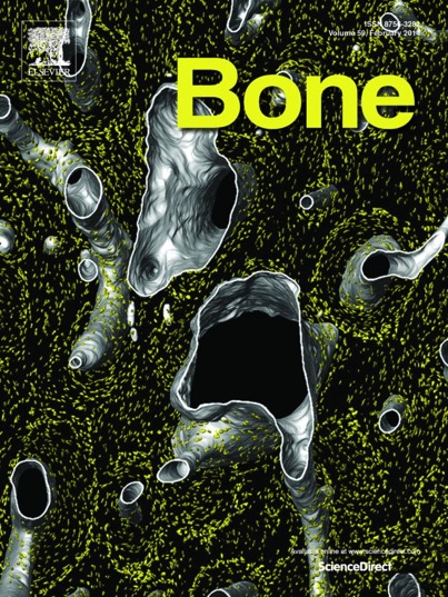
Figure 2 : Illustration of osteocyte lacunae (yellow) wuthin cortical bone around various osteons (grey) extracted from SR micro-CT images (voxel size 1.4 micrometre) – cover page of Bone, vol. 60, pp. 172-185, 03/2014
3D Phase Nano-CT imaging of the LCN
We also demonstrated the 3D imaging of the LCN by a new technique based on synchrotron phase nano CT (voxel size : 60 nm) [LANG-12]. These images provide not only the precise morphology of the porous network, but also information about collagen fibers [VARG-13c]. The technique developed here was applied to the study of the LCN in samples from patients with Biphosphonate Related Ostoenecrosis of the Jaw (BRONJ) in collaboration with the Humboldt University Berlin (PhD of B Hesse, co-supervision with K Raum) [HESS-14a][HESS-14b]
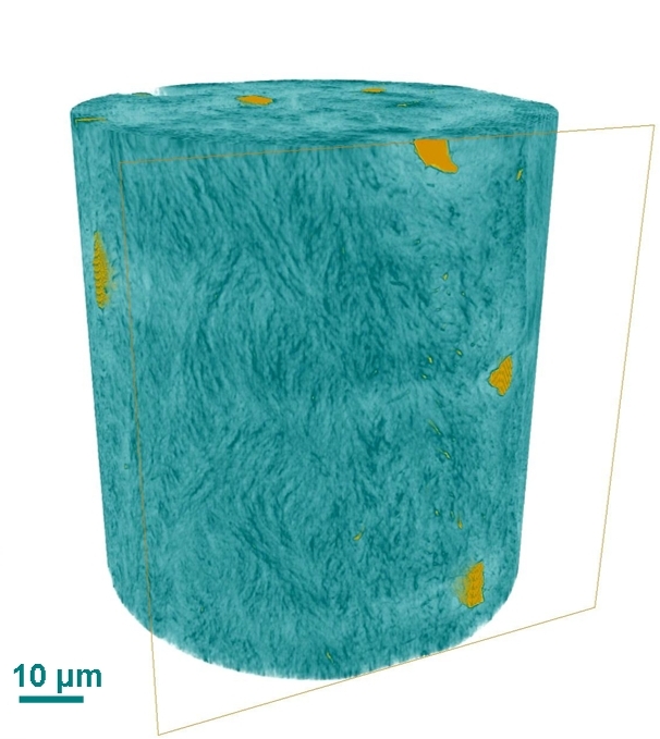
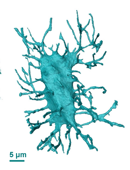
Figure 3 : Illustration of a single bone micro crack in 3D in trabecular bone after image segmentation : blue : micro-craks, pink : osteocyte lacunae and gray : surfaces of trabecular bone