Home

Main navigation

  • News
    • All news
    • Seminars
  • Presentation
    • CREATIS
    • Organigram
    • People directory
    • Staff
    • Contacts
    • Access
  • Research
    • Research teams
    • Transversal projects
    • Structuring projects
    • Imaging platform
    • Activity reports
    • Data information note
  • Contributions
    • Publications
    • Patents
    • Software
  • Studies & Training
    • Implications dans les formations
    • Doctoral Studies
  • Jobs Opportunities
  • French French
  • English English
Search API form
User account menu
  • Account
    • Log in

Breadcrumb

  1. Accueil
  2. 3D region growing segmentation

3D region growing segmentation

Purpose and Context

The goal of this work is to segment bone structures in three-dimensional [18]-NaF PET images.
The main difficulty in PET images processing lies in the high variations of the tracer uptake inside the organs. In the case of the [18]-NaF PET image, these variations may be also observed along the bone structures. To face to this problem, we propose to implement an adaptive region growing method with an automated initial seeds location.

Method

A region growing processing consists in merging all neighbouring pixels satisfying a homogeneity criterion starting from an initial set of seeds.
In an automated system the first step is the unsupervised location of the seeds GREN-05. This process is based on a priori knowledge about anatomical structures (skeleton, bladder, and kidneys) and their typical tracer uptake. This information is taken into account to compute special parameters used to locate the seeds.
In the region growing algorithm both local and global information is taken into account to the merging process GREN-06b. This approach is efficient to fit to the high variations of the tracer uptake.


Results

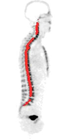
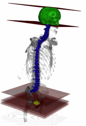
The methods were tested with 3D whole-body NaF PET images acquired on a high resolution PET scanner (CTI / Siemens ECAT EXACT HR+) in 3D mode protocol and reconstructed with AW-OSEM. The automated location of the initial set of seeds is shown in Fig. 1. Results of the adaptive region growing used to segment bladder and skull are displayed in Fig. 2.


b)Results of automated location of the seeds in the spine
c)  Results of automated location of the seeds in the head


Figure 1: Automated location of the seeds.
whole body NaF PET imageSeeds of the spine Seeds in the skull

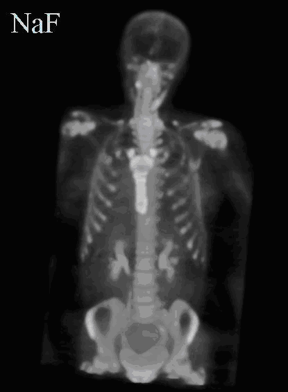
a) Maximum Intensity Projection of a NaF PET volume


Figure 2: Resulting automated segmentation of the head, the spine and the bladder by adaptive region growing.
Whole Segmentation

Barre liens pratiques

  • Authentication
  • Intranet
  • Rss feed
  • Creatis on Twitter
  • Webmail
Home

Footer menu

  • Contact
  • Map
  • Newsletter
  • Legal Notices