F. Peyrin
Purpose and context
Small animal imaging is raising increasing interest with the development of studies on animal models for therapeutics or genetics. Quantifying bone micro-architecture in mice has potentially many applications. Given the small size of bone structures in mice, it is necessary to work with high spatial resolution and a good signal-to-noise ratio.
Methods
We used synchrotron micro-CT to quantify micro-architecture in a mice model of osteoporosis by hindlimb suspension developed at the LBTO (Inserm E366 Saint-Etienne). For this purpose, we developed an automatic image analysis technique to identify and quantify the trabecular and cortical envelopes of the femoral metaphysis [Martin-Badosa et al. Comput Med Imag Graphics 2003].
|
a) |
b) |
c) |
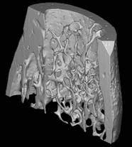
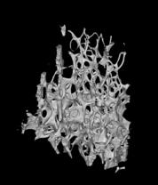
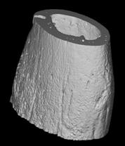
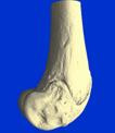
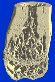
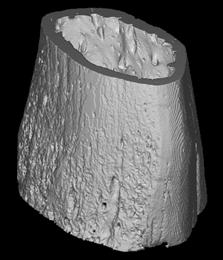
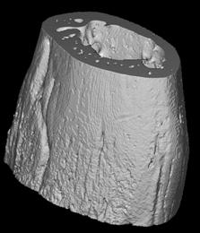
3D display of femoral mice bone metaphysis imaged by synchrotron micro-CT (voxel size : 6.7 µm): a) volume of interest (half) which is splitted in b) trabecular and c) cortical volumes of interest.
Results
The 3D images of the excised mice femurs, obtained with a voxel size of 6 µm, give a good rendering of the bone trabeculae in the mice, the dimensions of which were found around 20-30 µm.
|
a) |
b) |
c) |
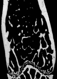
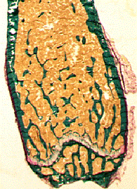
Femoral mice bone metaphysic imaged by synchrotron micro-CT (voxel size : 6.7µm) :a) longitudinal slice, b) comparison to a typical longitudinal slice obtained by histology (from LBTO), c) 3D display.
The 3D SR-micro-CT images made it possible to quantify significant differences in architecture and mineralization between two strains of mice (C3H/HeJ@Ico and C57BL/6J@Ico, referred to here as C3H and B6) [Martin-Badosa et al., Radiology 2003]. In addition, these two strains did not have the same response to the effects of suspension, with a loss of bone tissue accompanied by thinning of the trabeculae observed in the B6 strain [Martin-Badosa et al., Comput Med Imag Graphics 2003].
|
a) |
b) |
Synchrotron micro-CT images illustrating the differences in the cortical bone envelope on two strains of mice : a) C3H (thick cortex), b) B6 (thin cortex)
In order to do longitudinal follow-up, we recently showed the feasibility of imaging mice femurs in vivo on the synchrotron micro-CT system. The initial quantitative results on the two strains of mice (C3H and B6) imaged in vivo (voxel size : 10 µm) showed the expected significant differences in micro-architecture. As compared to standard micro-CT, this technique has the advantage to provide high quality images in very short acquisition times ( < 5 min) for doses of about the same order of magnitude [Bayat et al, Nucl Instr Meth A 2005]. Another perspective concerns the analysis of data acquired in mice fetuses. transgenic mice fetuses In a study, carried out with the group of S. Majumdar (San Francisco, Dept of Radiology, USA), transgenic mice fetuses were imaged at very high spatial resolution (~ 1 µm) to evaluate the role of Insulin like growth factor-I (IGF-I) on bone formation [Burghardt et al., Bone 2007].
References
[1]- MARTÍN-BADOSA E., ELMOUTAOUAKKIL A., NUZZO S., AMBLARD D., VICO L., PEYRIN F., A method for the automatic characterization of bone architecture in 3D mice microtomographic images, Computerized Medical Imaging and Graphics, Nov. 2003, vol 27, n°6, p. 447-458.
[2]- MARTÍN-BADOSA E., AMBLARD D., NUZZO S., ELMOUTAOUAKKIL A., VICO L., PEYRIN F., Excised bone structures in mice: imaging at three-dimensional synchrotron radiation micro CT, Radiology, Dec 2003, vol 229, n° 3, p. 921-928.
[3]- BAYAT S, APOSTOL L, BOLLER E, BROCHARD T, PEYRIN F, In vivo imaging of bone micro-architecture in mice with 3D synchrotron radiation microtomography, Nucl. Instr. and Meth. A, (Elsevier), 2005, vol. 548, p. 247-252.
[4]- BURGHARDT A, WANG Y, ELALIEH H, THIBAULT X, BIKLE D, PEYRIN F, MAJUMDAR S, Evaluation of fetal bone structure and mineralization in IGF-I deficient mice using synchrotron radiation microtomography and Fourier transform infrared spectroscopy, Bone, 2007 Jan, vol 40, n° 1, p. 160-168. Collaborations and acknowledgements This work was performed in collaboration with ESRF (Grenoble), LBTO, Inserm E366 Saint-Etienne, and University of San Francisco (USA).