This reasearch theme is conducted through a collaboration with the LabTau and in the framework of the LabEx CeLyA. It leads to the co-supervision of three doctoral projects in the context of cavitation-based therapy where a high-intensity focused ultrasound transducer is used to create cavitation bubbles that induce mechanical damage. The created nuclei must be imaged in order to control and monitor the therapeutic device. To evaluate the position and movement of the bubbles, passive imaging was proposed. Indeed, during therapeutic pulses, the energy present in the medium is too high for active ultrasound imaging. The developed experimental platform includes an imaging system that is triggered to visualize the bubbles during cavitation pulses from the received raw signals. Original receiving beamforming algorithms have been proposed to create cavitation maps with a high spatial resolution. First of all, the use of phase and sign coherence factors made it possible to validate all the configuration and post-processing sequences [Boulos-2018-IEEE Transactions on Ultrasonics, Ferroelectrics and Frequency Control]. It also lead to validation of the measure accuracy in in vitro fantom.
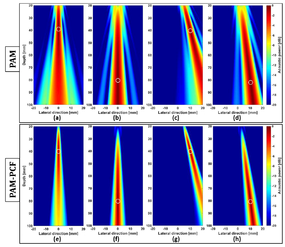
From simulation dataset of a point source, images reconstructed using (a–d) PAM and (e–h) PAM-PCF. In each subplot, the theoretical position of the point source is marked by a white circle, with the (lateral; depth) coordinates in mm from left to right: (0; 40), (0; 80), (10; 40), and (10; 80).
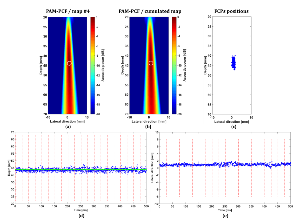
Working in agar gel and using LA523 imaging probe with CI=20. (a) The cavitation map reconstructed with the PAM-PCF method for the 4th passive acquisition frame. (b) Cumulated map along 1500 maps. The white circle corresponds to the FCP localization. (c) The FCP 2D positions. The FCP localization temporal profile along 500 ms of sonication for both (d) depth and (e) lateral imaging direction. The horizontal green line represents the FCP theoretical depth reference and the vertical red dotted line corresponds to the start of each sonication pulse.
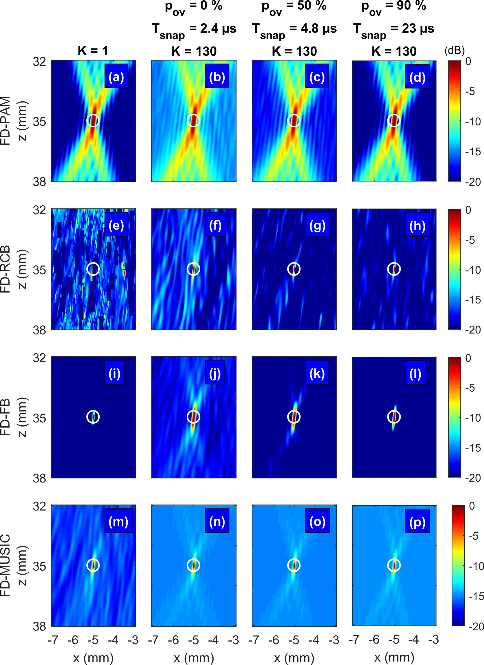
Then, specific adaptive beamformers were proposed in the Fourier domain to improve both resolution and contrast. These beamformers (minimum variance, functional beamforming) are based on a robust estimate of the cross-spectra of the received signals, which is not the case in the literature, and which was successfully addressed. The great advantage of the proposed beamformers is that they can be used in both passive and active acquisition [Polichetti-2018-Applied Sciences].