NEWS
The platform's ultrasound modality is now equipped with a matrix probe compatible with the ultrasound scanners Verasonics : 32 x 32 elements - central frequency : 3 MHz - produced by Vermon
DESCRIPTION OF ULTRASOUND PLATFORM
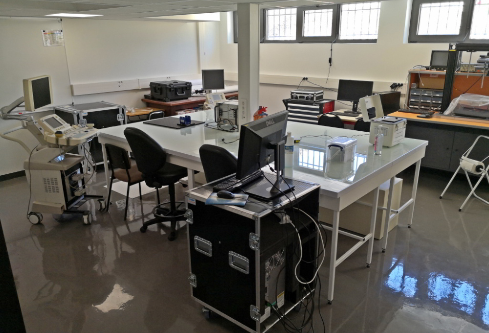
The Lyon-Tech PILoTplatform has a part dedicated to the ultrasound imaging, patircularly developped and enhanced by tools or advanced solutions by the ultrasound imaging team og CREATIS, composed by :
- Seven ultrasound scanner :
- whose 3 clinical with research options of Ultrasonix brand
- with 4 of research kind :
- 2 Ula-Op, developed by the university of Florence, are used in research for the development of specific methode in antenna treatment (Zahnd_2015).
- 2 Vantages from Verasonics'company : control of 512 synchronised channels and so to use matrix probes, especially one available on the platforme of 32 x32 elements, in order to, by example, verify the sparse array's (Roux 2018)
- Phantoms : we have tools allowing the realization and characterization ( Duboeuf_2009, Duboeuf_2007) of gels mimicking the biological tissue of arterial wall (Brusseau_2001), of prostate tissue (Boutey_2009, Vray_2009 brevet) and cancerous (Brusseau_2007, Brusseau_2008). A lot of brand phantoms are avalaible in the platform.
- An automatic displacment banch
- A group of electronical devices (low frequency generator, Lecroy's scope, transmitter/receiver mono-channel, hydrophone with a band-pass from 1 to 30 MHz, piezo-électrical element, etc...).
- Bi-modality : in the platform, is possible to have access to the bi-modality : ultrasound/optic (Vallet_2017) et ultrasound/MRI
-The echographs
The three echographs of the Ultrasonix'brand (RP, MDP and Touch, the last bought in 2014), allow the record of ultrasound waves and the programmation of the most of the echograph's components. This characteristic is fundamental for the modification and the analysis of raw signals, allowing the improvment and the creation of news methodes of diagnosis like elastography (Brusseau_2014). Is it possible to test prototype of probe (Brusseau_2017). Volumes acquired by a 3D probe are used in the study of needle tracking (Zhao_2016).
The Ula-Op allow, in addition to functionalities near of Ultrasonix, the transmission of arbitrary signals. They can also be used for the le development of new harmonic methodes of imaging (Toulemonde_2015, Lin_2013). Specific strategies of emission/receiving are programmables to test, by example, algorithmes of compressed sensing (Lorintiu_2016).
The two Vantage, like the Ula-op, are completely computable and provide test of some new imaging sequences such as divergents waves (Zhang_2016). The access of pre-beamforming data gives the possibility to test new reconstruction methods like neuronal network (Gasse_2017). Otherwise, a power module allows to transmit long pulses for, especially, elastography by shear waves.
The seven echographs of the laboratory are real technologic platforms of research in ultrasound imaging. Their functionality in research mode, installed by the manufacturer, provides access to signals at all stages of processing. The RF, Post scan, Pre-scan signals can be saved. The library proposed by the manufacturer provides :
- The integration in real-time of image processing methods.
- The connexion and the management of prototype probe.
- access to raw signals to test new methods of beamforming.
- The synchronization with external manipulations from a distant office. control of imaging aquisition.
- The choice of the emitted signals with a programmable generator.
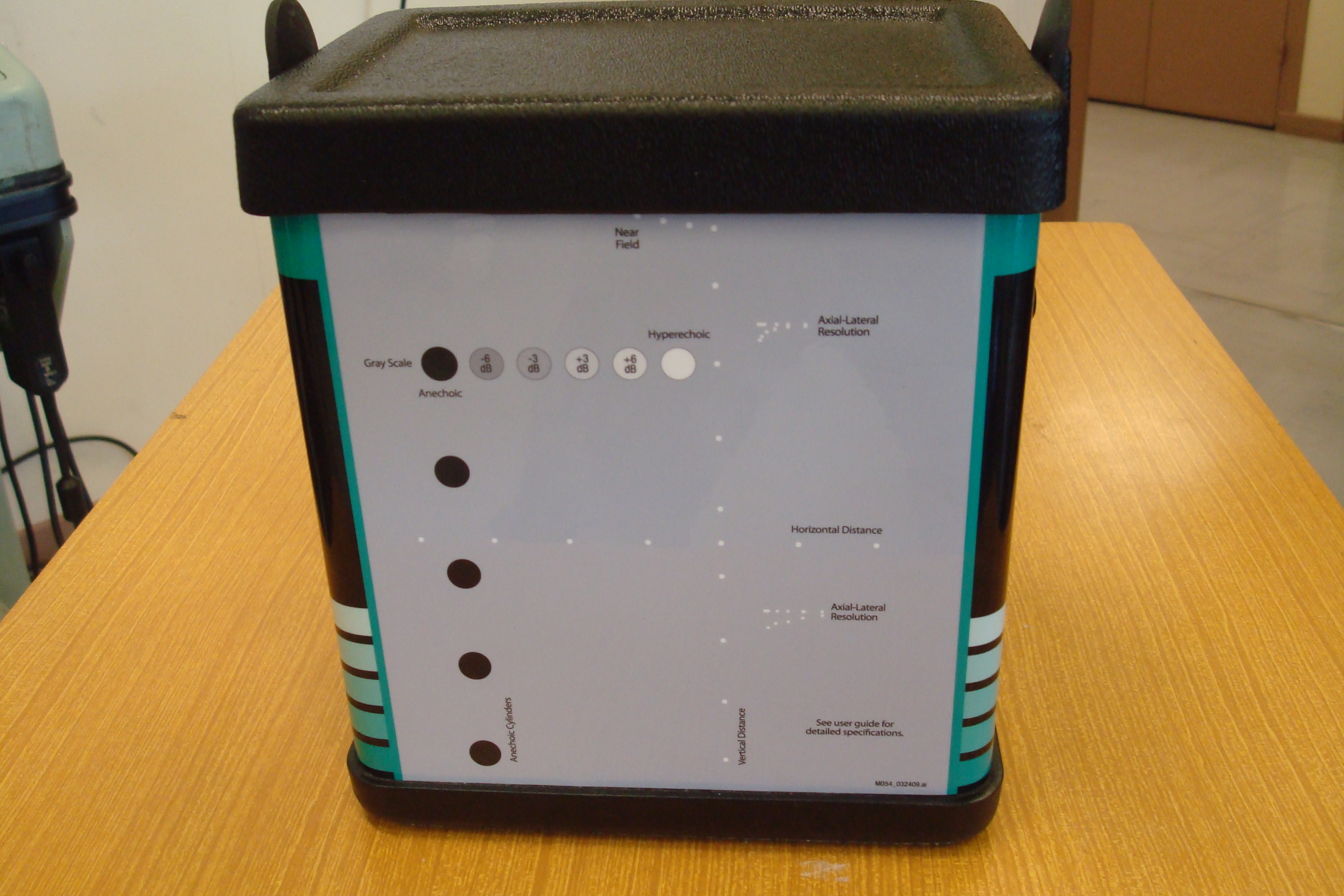
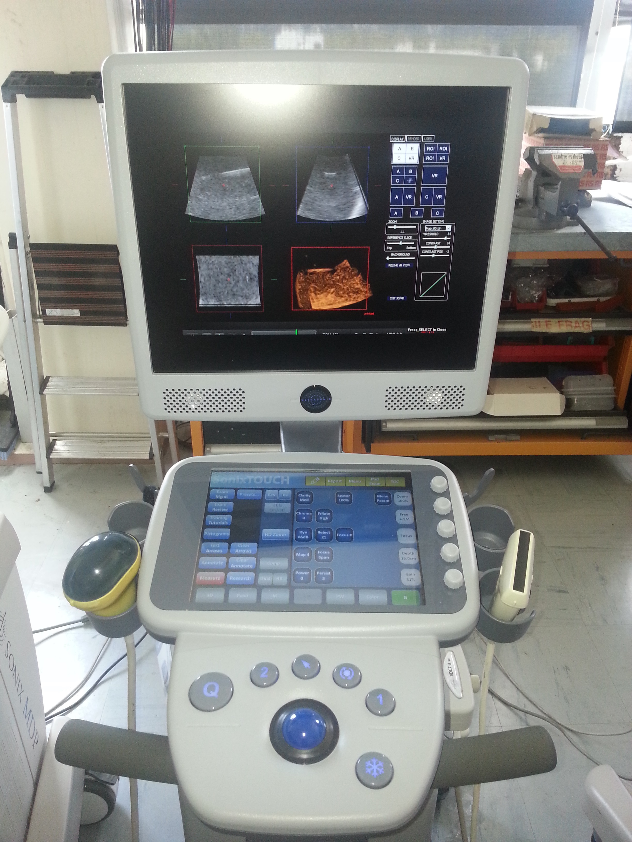 | Ultrasound system Ultrasonix : Touch (on the picture), MDP et RP Characteristic technic Studies completed : Elastography (Elavisu) Propello RANSAC Available probes : linear, 3D, cardiac, CMUT, multi-plane | ||
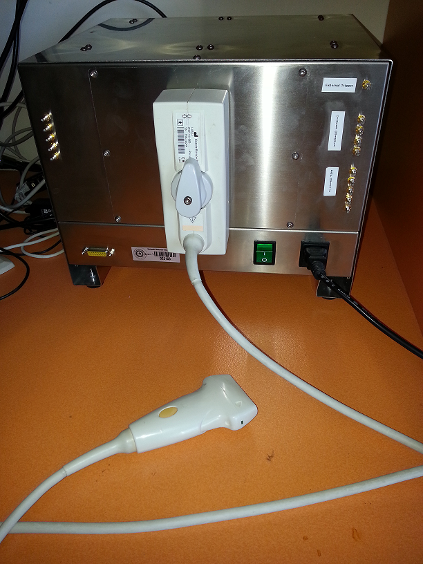 | Ula-Op (Ultrasound Advanced Open PLatform) are systems developped by the MSD-Lab of Florence's University. The first has 64 channels and the second has 256 channels programmables in transmission/reception. These ultrasound scanner allows to transmit signals of arbitrary shapes thanks to their programmable generators. Available probes : linear, cardiac, matrix, CMUT | ||
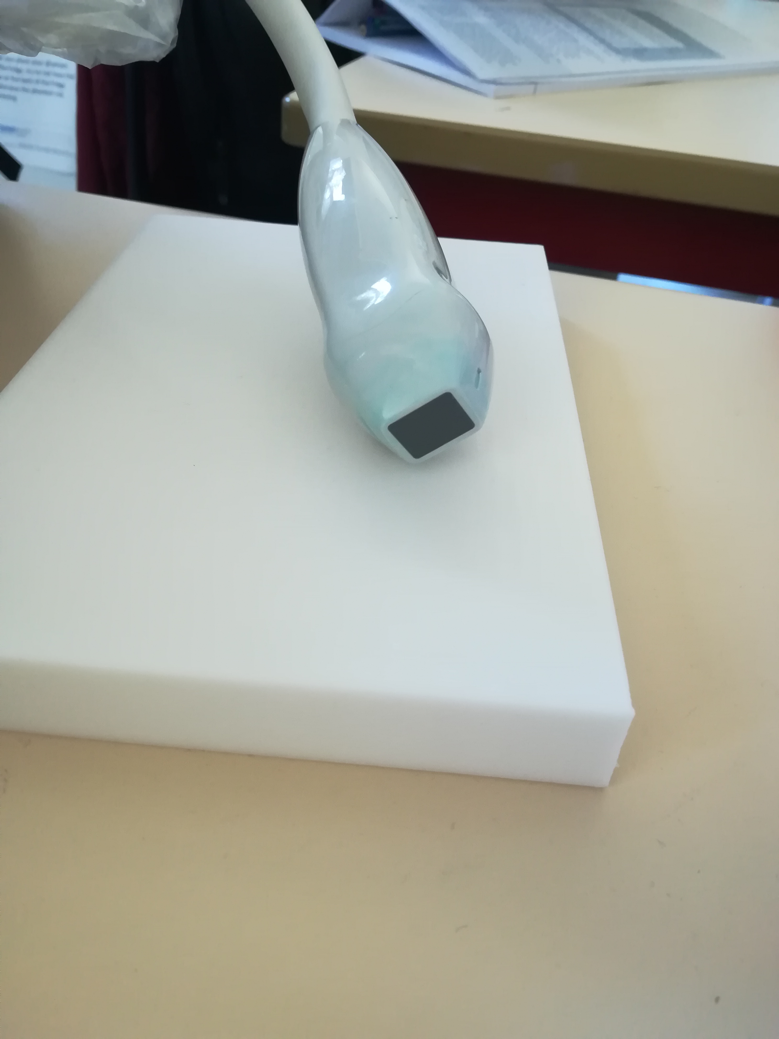
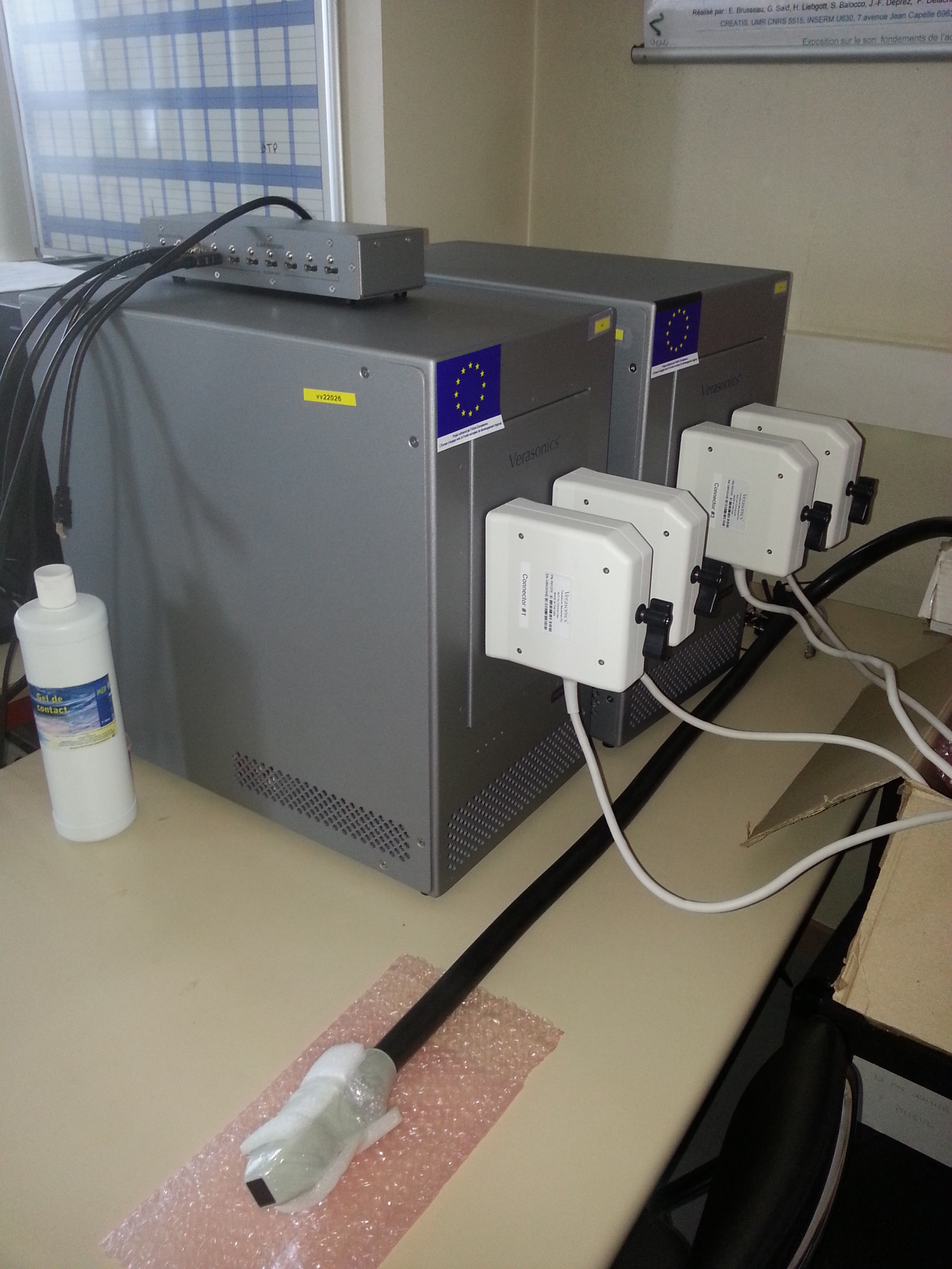 | 2 Vantage systems commercialized by the american society Verasonic. Each has 256 programmables channels in transmission/reception. Being synchronizable, it is possible to have 512 parallel way. A power module allow especially to realize elastography by shear waves. The Verasonics system was acquired thanks to the program FEDER Saint-Etienne and Loire General Council in the framework of the project SonoCardioProtection supervised by Pierre Croisille. Probes availables : linears, cardiacs, convex. A matrix probe, 32 x 32, so 1024 channels, visible on the image, is acquired with the labtau, INSERM U1032. | ||
Concretes examples of realisation :
- Elastography studies
The ultrasound elastography is an imaging method, who, from digital treatment of ultrasound radio frequency data, gives information on the mechanical properties local of a medium.
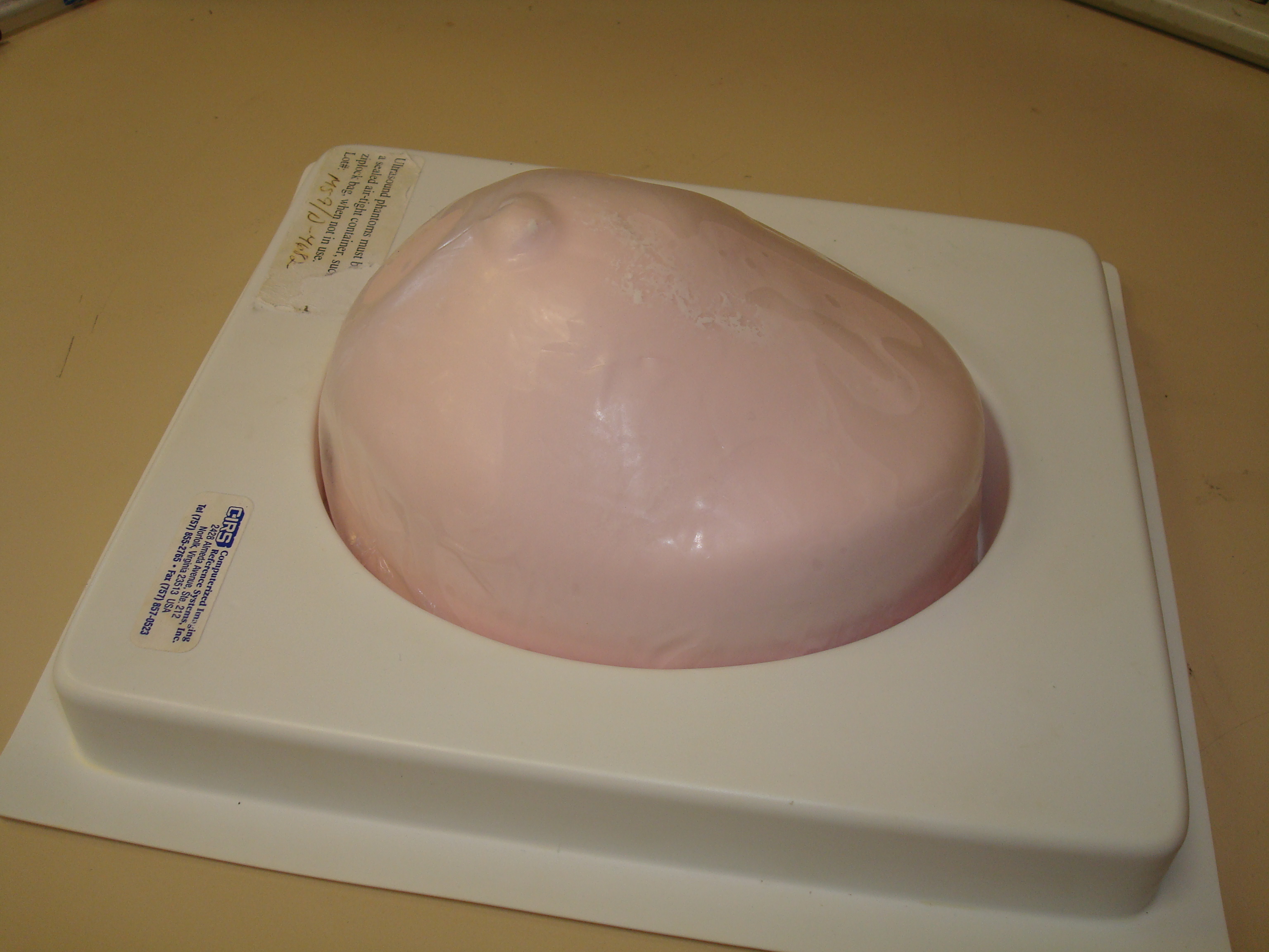
Illustration with the breast phantom (presented below) :


| Echography of the phantom on an inclusion that has an elasticity module taller the the medium. This inclusion is less visible on the ultrasound image | Elastography result showing the deformation of the medium during the compression. The inclusion is clearly visible (at the center of the image) |
- Imagerie non-linéaire
improvment of the Contrast Tissue ratio (central image) by using a pulse inversion method (on flow phantom, explained below)

-Les fantômes
-"Hand made"
The platform knows how to realize phantoms (test object mimicking biological soft tissue for the ultrasound characteristics).
These phantoms could be characterized by them mechanical and ultrasound properties with tools available in the laboratory (tank, hydrophone, mono_transducers ...)
 | Réalisation de fantômes en Agar-Agar, Gélatine et Polyvinyl Alcool en fonction de la nécessité de l'étude. |
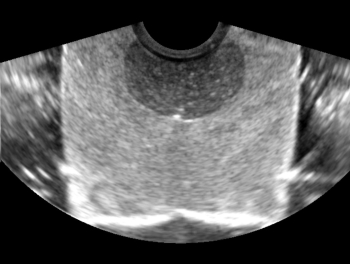
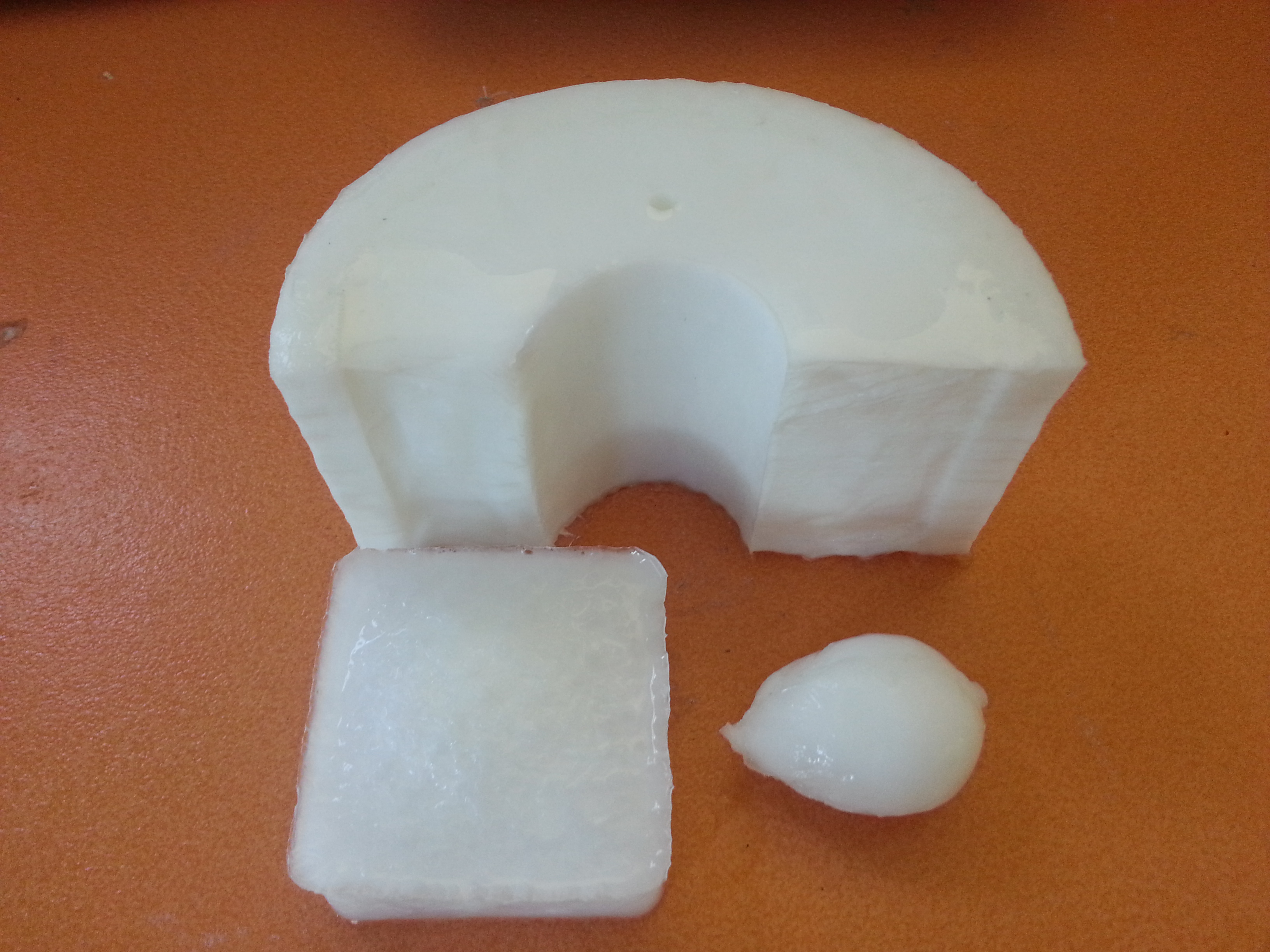 | 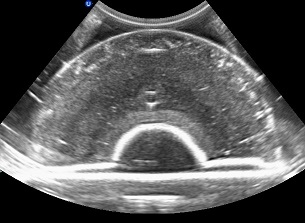 | Exemples de fantômes en Polyvinyl-Alcool de géométrie différentes. A droite est présentée l'image échographique du fantôme en demi-cercle. |
- les fantômes commerciaux
Le plateau dispose également de plusieurs fantômes achetés à des sociétés spécialisées :
 | 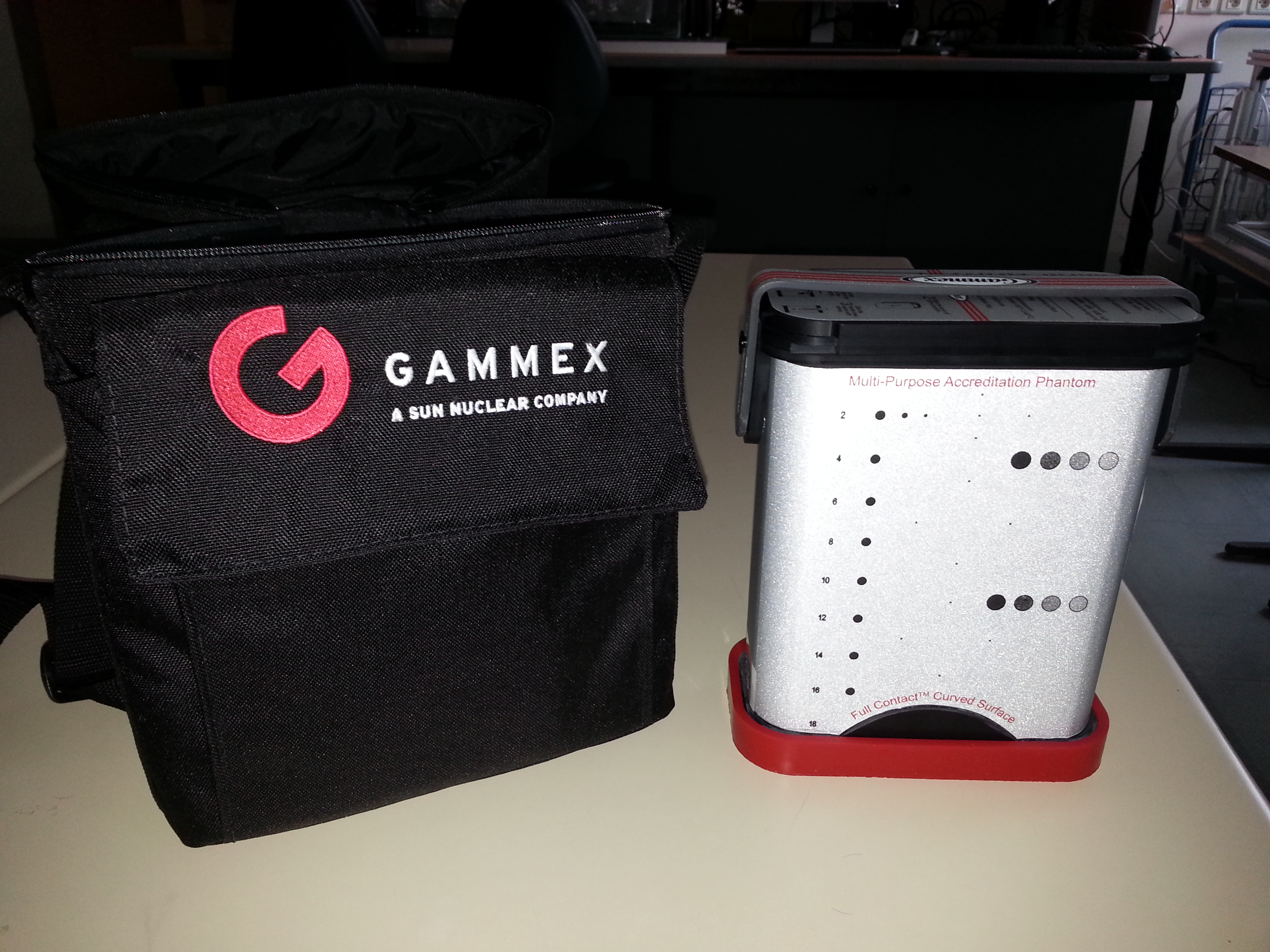 | De gauche à droite : - fantôme de calibration générale de marque CIRS modèle 054GS - fantôme de calibration générale de marque Gammex modèle 410 SCG0.5 |
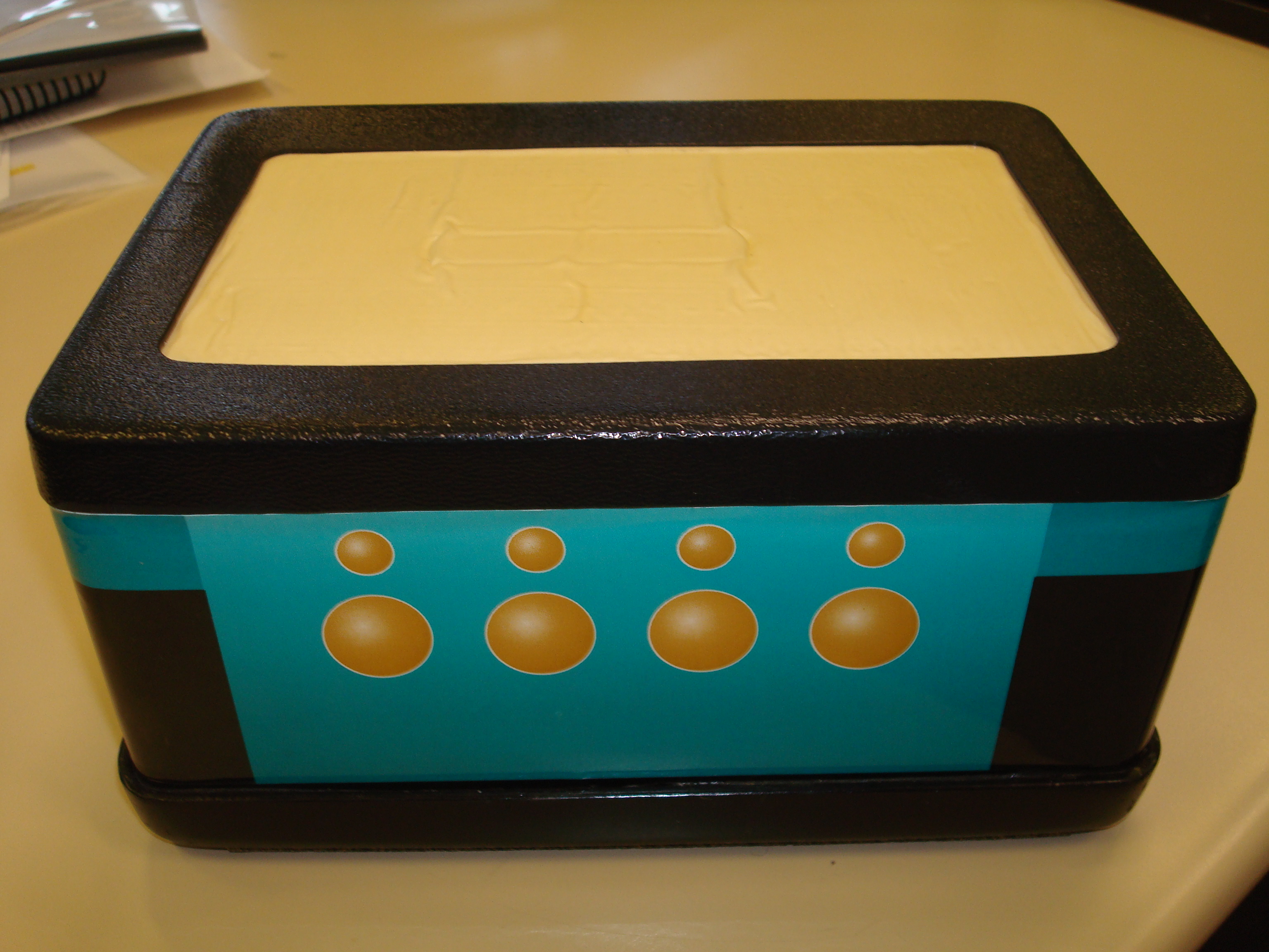 |  | Fantômes pour l'élastographie de marque CIRS (de gauche à droite) : - modèle 049 - modèle 059 |
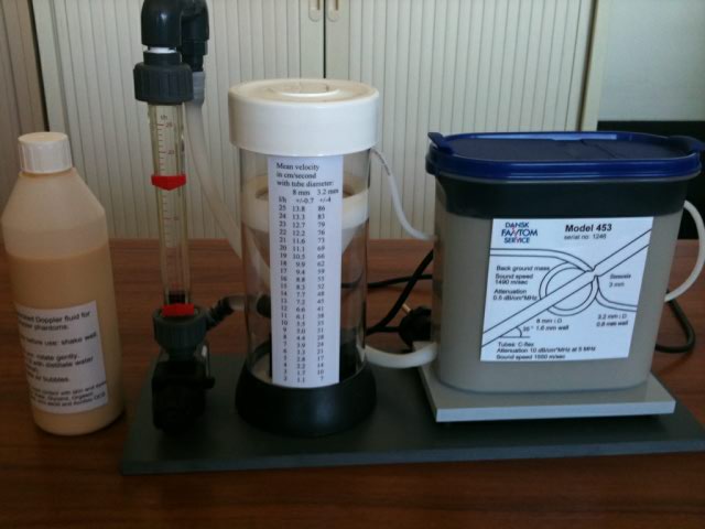 | 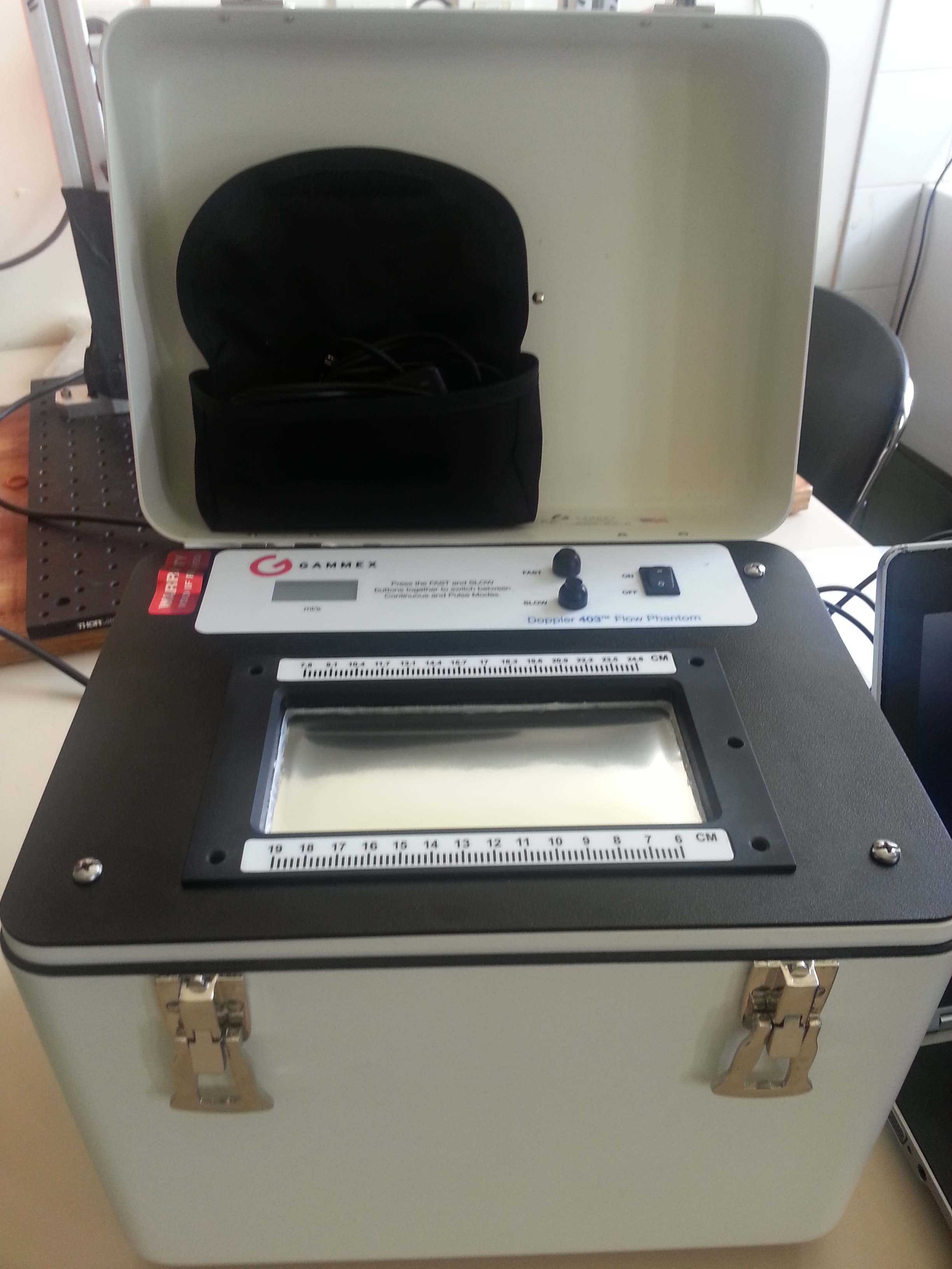 | Flow phantom for doppler imaging or others flow imaginig (from left to right) : - Doppler phantom from Dansk Fantom model 453 - Doppler phantom - continuous and pulsed flow from Gammex model Doppler 403 |
- Phantoms from collaboration
We have the luck to have on our platforme a vortex'phantom from the Leeds Test Objects' Company :
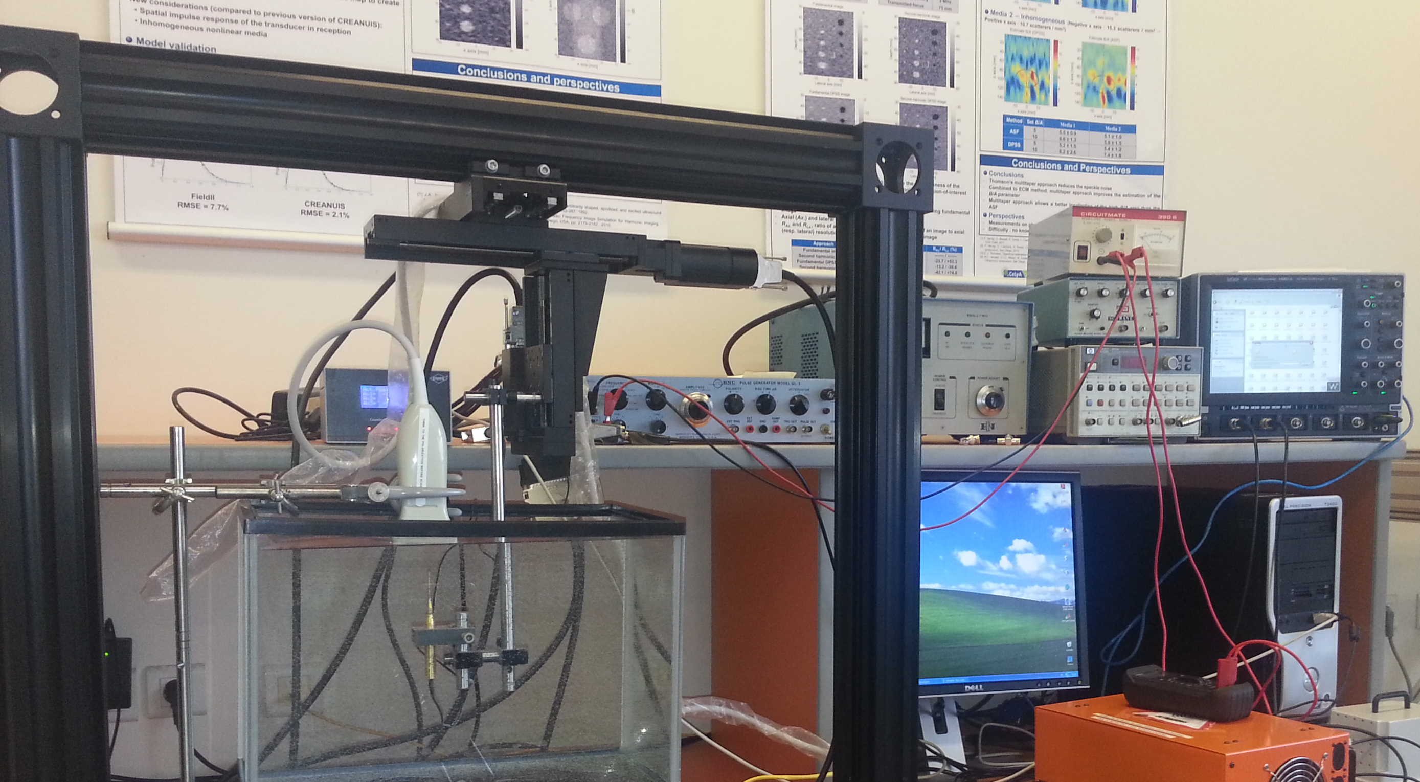
Il permet le déplacement automatisé axial, latéral et transversal des sondes échographiques utilisées au moment des acquisitions. Les algorithmes, conçus et adaptés pour et par les chercheurs de l'équipe, permettent une souplesse d'utilisation. Des séquences d'acquisition en quasi-statique, sous déplacement controlé et connu, permettent de valider des méthodes d'estimation de mouvement. Une application Labview permet de caractériser des sondes ultrasonores. Ce banc a récemment été complété par deux axes en rotation qui peuvent se fixer avec les autres axes linéaires.
|
Caractéristiques techniques du banc de mesure ultrasonore :
|
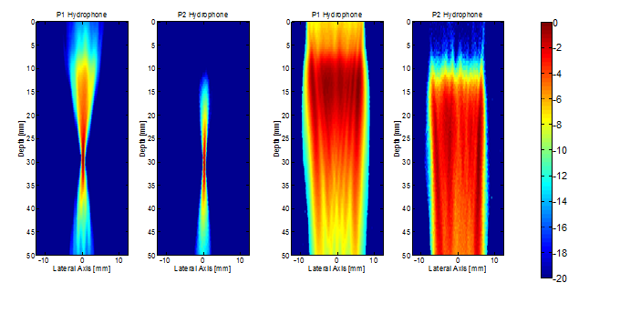
Un exemple de résultat :
Valeur du fondamental et du second harmonique du champ de pression d'une sonde linéaire en focalisation et en onde plane
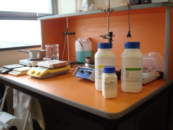
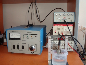
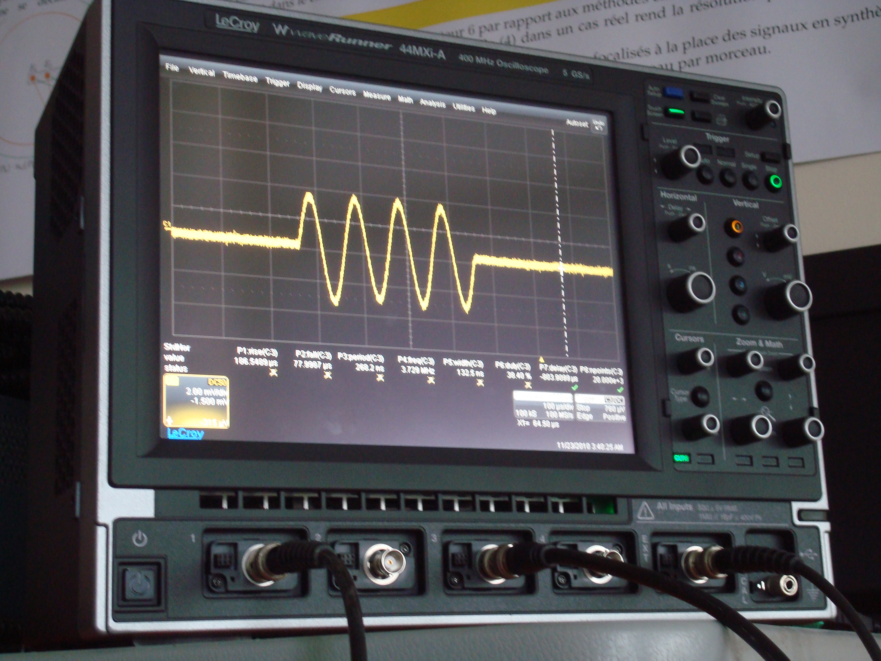
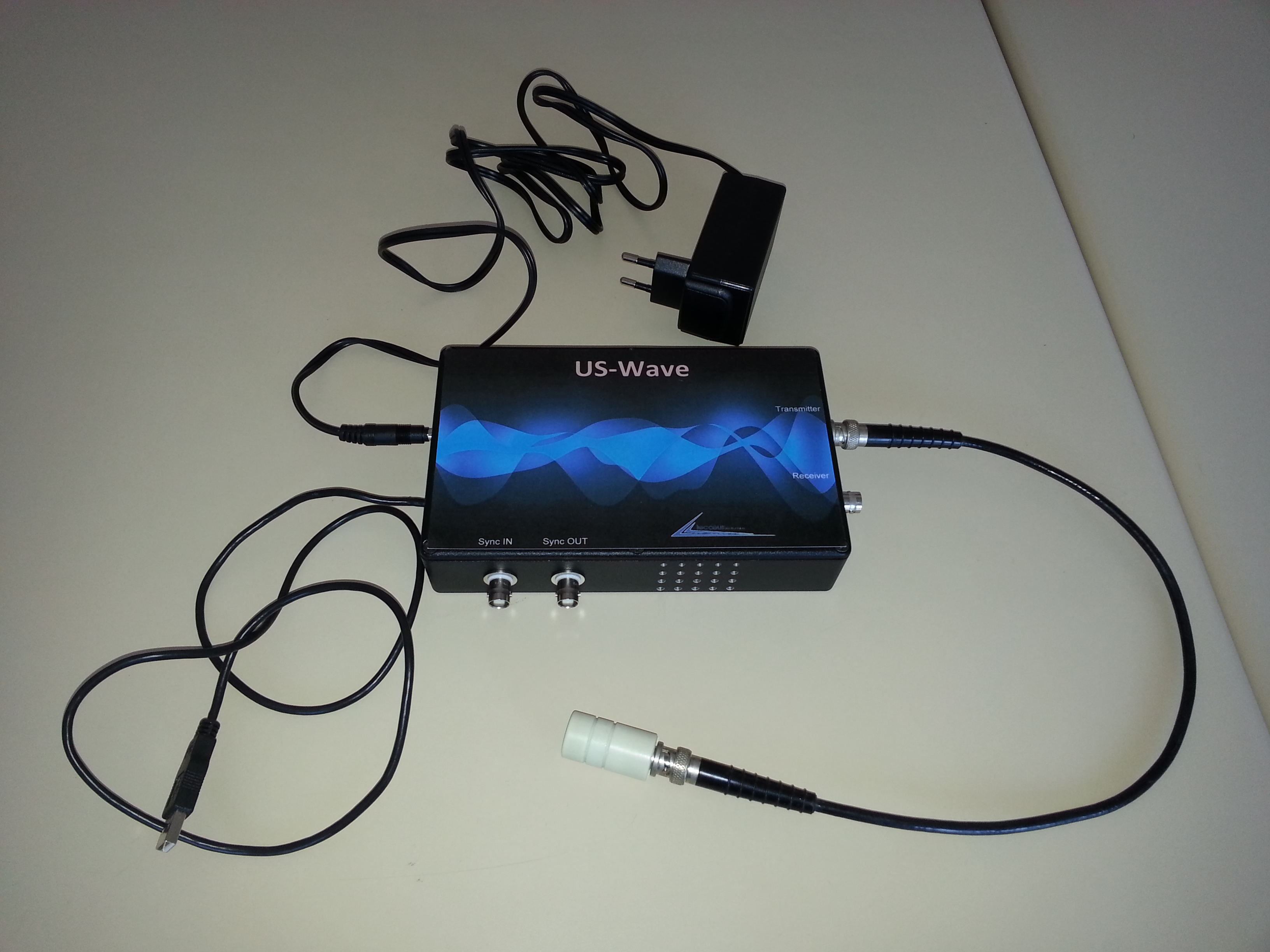
-Les nombreux instruments électroniques
Synthèse et lecture de signaux électroniques sont nécessaires aux études réalisées. Ceci est permis notamment par un oscilloscope LECROY (Oscilloscope WaveRunner Xi-A/MXi-A), des générateurs de fonction, des amplificateurs de gain. Un émetteur / récepteur monovoie US wave (de la société Lecoeur électronique) est pilotable sous Matlab, ainsi qu'un choix important d'éléments mono-transducteur (dernière acquisition en date : 2 mono-transducteurs de 5 MHz de marque Sofranel).
- BI-MODALITE
Ultrasons / Optique
Salle photoacoustique
Au sein de la plateforme nous possédons une salle permettant de réaliser de la photo-acoustique :
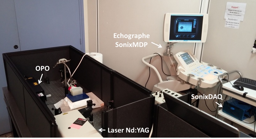
| Caractéristiques du laser de pompe Nd:YAG | Caractéristiques de l'OPO (Oscillateur Paramétrique Optique) | Echographes: |
- λ = 355 nm - Fréquence de répétition = 10 Hz - tp = 5 – 8 ns - d ~ 8 mm - E < 200 mJ / impulsion | - 410 nm < λ < 2550 nm - Fréquence de répétition = 10 Hz - tp = 3 – 7 ns - d < 5,5 mm - E < 8 mJ / impulsion | Les acquisitions photoacoustiques peuvent être réalisées avec nos échogarphes Ultrasonix MDP + DAQ ou avec le Ula-Op |
Pour en savoir plus sur cette modalité et les résultats obtenus : cliquer ici
Fantôme bi-modaux
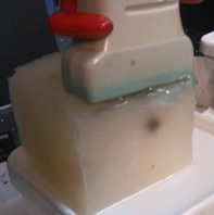
 |  | Exemple de fantôme en Polyvinyl-Alcool modélisant les trois couches d'un tissu prostatique (collaboration avec le CEA-Leti.) |
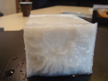
 | 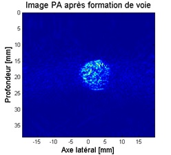 | Exemple de fnatôme réalisés pour la photoacoustique : une inclusion sphérique teintée de noir dans un milieu environnant en PVA et sa visualisation en photoacoustique |
Ultrasons / IRM
Cette bi-modalité est exploitée par le biais de l'élastographie. Nous fabriquons sur le plateau des fantômes en plastisol. Ils sont ensuite passé dans l'IRM de la plateforme pour acquérir des données ERM :
Fantôme bi-modaux
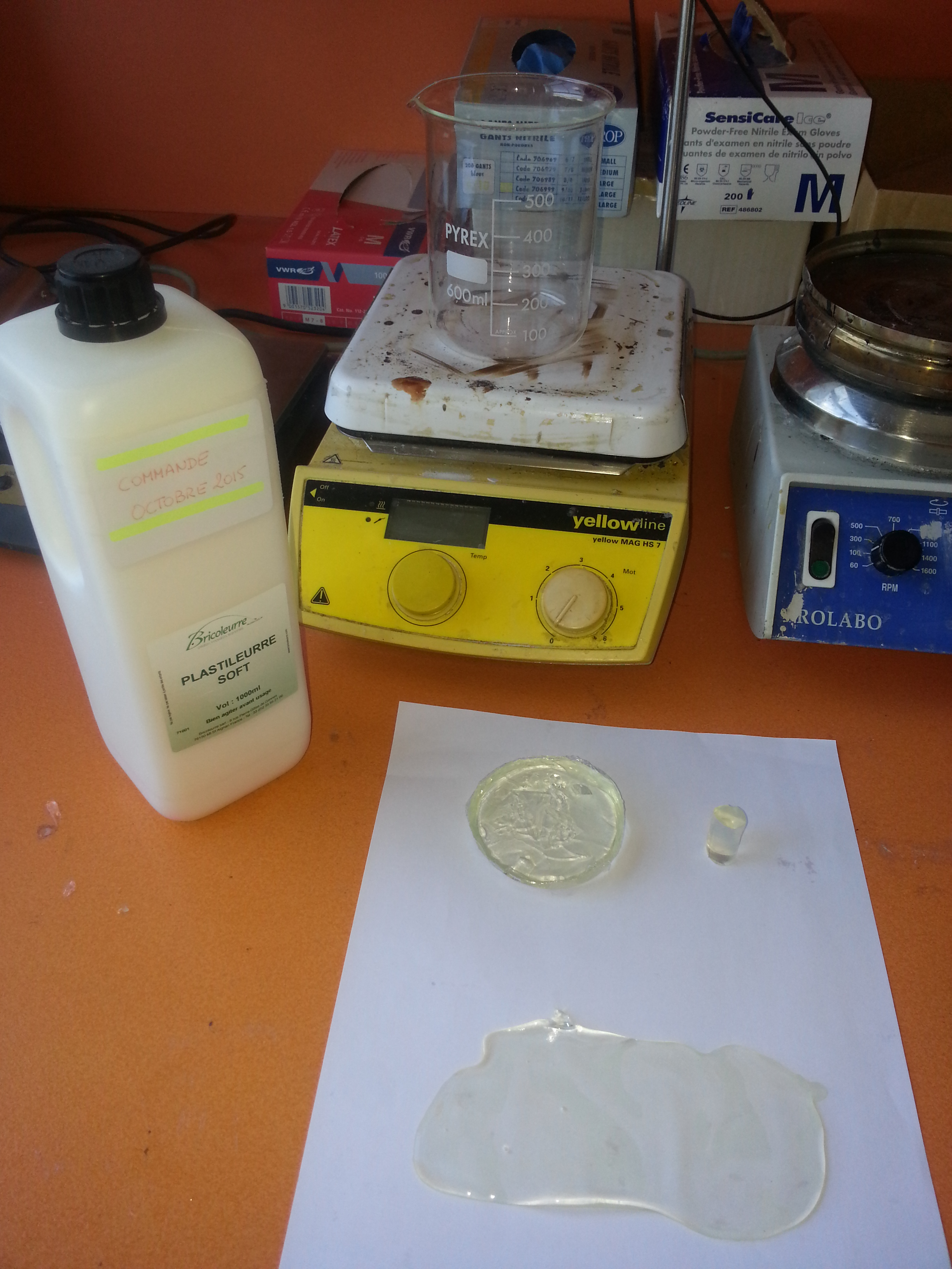 |  | Réalisation de fantômes en plastisol et visualisation ERM de la propogation d'onde de cisaillement |
Pour en savoir plus sur cette modalité et les résultats obtenus : cliquer ici
QUI CONTACTER
Tél : +33472431887, mail : Adeline.Bernard@creatis.insa-lyon.fr
Tél : +33472431887, mail : Denis.Grenier@creatis.insa-lyon.fr