Purpose and context
Microdamages accumulating in bone tissue due to mechanical constraints associated to daily life may affect the integrity of bone. However, they are also supposed to have a strong role in the reparation processes and to trigger bone remodeling. Linear microcracks are considered as thin plane defects (opening ~1 µm). Their analysis remains difficult and is mainly based on stained histological slices, thus providing only partial results.
Methods
We demonstrated the possibility of quantifying micco-cracks in 3D by using synchrotron radiation (SR) micro-CT at the micrometer scale and adapted 3D image analysis methods.
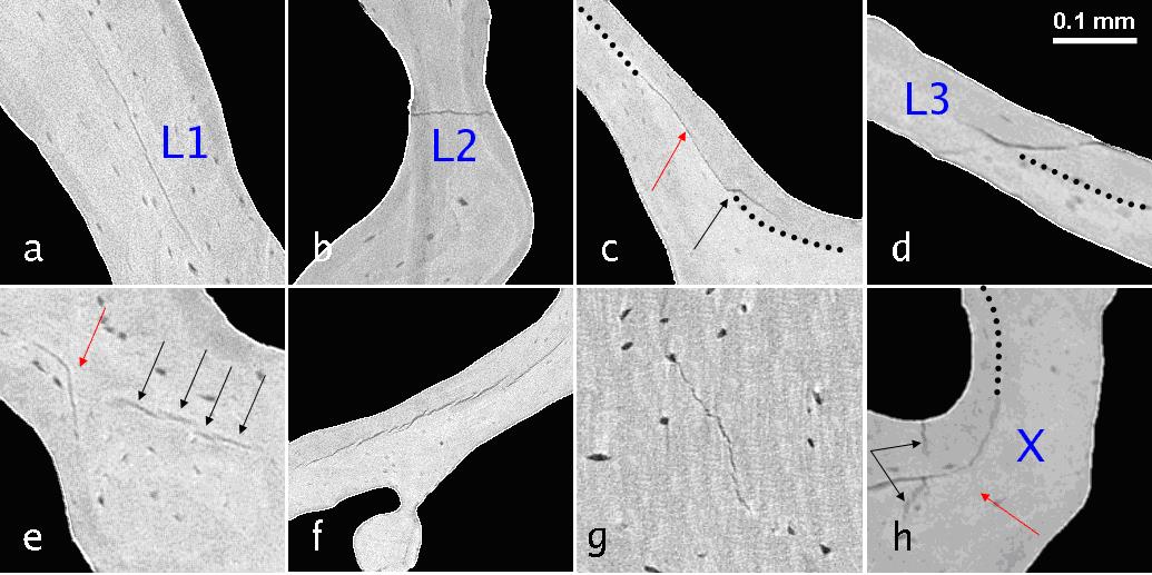
Figure 1 : Illustration of different types of bone micro cracks in trabecular bone
Results
In the PhD of Aymeric Larrue, we developed a method based on 3D SR micro-CT at the micrometric scale to study microcracks in human trabecular bone (collaboration with L. Vico, LBTO, U1059, St-Etienne). After the optimization of image acquisition, different types of micro-cracks could be observed in the SR micro- CT slices. We proposed a specific method for the segmentation of micro-cracks using a non linear filtering based on a 3D steerable filter [LARR-11]. Analysis of cracks in cortical bone was performed in collaboration with the LIP (P Laugier, Paris) in order to validate a nonlinear acoustic method to assess microcracks in vivo [HAUP-12] [HAUP-14]. Further studies are in progress with the group of P Zysset (Bern, Switzerland) and the codes developed within the PhD of A Larrue are being used on a large series of samples to segment microcracks generated by different types of biomechanical constraints.

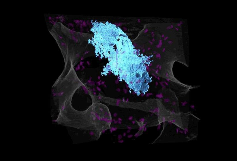
Figure 2 : Illustration of a single bone micro crack in 3D in trabecular bone after image segmentation : blue : micro-craks, pink : osteocyte lacunae and gray : surfaces of trabecular bone