Quasi-static ultrasound (US) elastography and magnetic resonance (MR) elastography are two techniques available in our laboratory, developed by the Ultrasound Imaging Team and the NMR and optics Imaging Team, respectively. Our objectives are to analyze which approach better suits for a specific medical application and to investigate the potential of combining the two techniques. Deep assessment and comparison of US and MR elastography have already been initiated

Fig. 1: Description of the CIRS phantom, model 049. (a) Phantom picture, (b) Top view of the phantom and (c) right end view. This test-object contains eight spheres of different size (10 and 20 mm), location and mechanical properties. The background material has a Young’s modulus of 29 kPa, whereas spheres have a Young’s modulus of 6 kPa (in blue), 17 kPa (in orange), 54 kPa (in green) and 62 kPa (in yellow).

Fig. 2 (a) Amplitude, (b) Shear wave visualization, and (c) Shear modulus cartography (kPa) at the S1, S2, S3 and S4 inclusion slice position.
The MRE measures realized (3µ=6kPa, 15kPa, 37kPa and 41kPa respectively) are in good concordance with the inclusion characteristics.
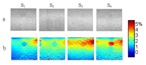
Fig. 3 (a) B-mode images and (b) Strain images (in %) obtained for the four phantom locations scanned S1, S2, S3 and S4.
US-E puts in relief inclusion positions and provides information on strain over time.
[1] D. Grenier, E. Brusseau, H. Liebgott, F. Duboeuf, F. Pilleul, and O. Beuf. MR-Elastography versus quasi-static US-Elastography. In ESMRMB, Antalya,Turkey, 2009.