F. Peyrin
Purpose and context
Due to the physical properties of x-ray beams extracted from synchrotron sources, synchrotron micro-CT is as a reference method for evaluating new imaging techniques for the investigation of bone micro-architecture. In particular, it was used to evaluate the precision of a MRI micro-imaging setup and a commerialized micro-CT system. Methods MRI micro-imaging was done at the U2R2M in Orsay (G. Guillot, J. Bittoun, CNRS UMR), on a high field imager (7T) (isotropic voxel: 80 µm). The aim of the study was to evaluate the incidence of MRI artefacts on the quantification of the trabecular network. The originality of the study was to develop a method allowing to match the images in the two modalities in order to compare the parameters in exactly the same region of interest (ROI). The results of MRI were found to correlate well with the measurements from the micro-CT but with a systematic bias in the values
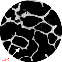
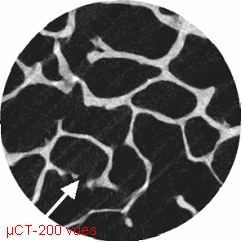
Standard micro-CT imaging was done in Orléans (C.L. Benhamou, C. Chappard, Inserm U658). Samples of sub-chondral bone taken from patients with arthrosis and osteoporosis were imaged at 10 µm both in micro-CT at the ESRF (900 projections) and on the Skyscan 1072 system for different numbers of projections (200, 400 and 800). Micro-architecture parameters were measured on similar ROI on thresholded volumes. The results show a good correlation between the measurements when standard micro-CT acquisition uses 800 projections which degrades when the number of projections decreases
Results
|
(a) |
(b) |
Comparison of 2D matched slices extracted from 3D micro-CT volumes (voxel size 10 µm) : a) acquisition from synchrotron radiation micro-CT (900 projections) b). acquisition from commercialized micro-CT (200 projections) showing blurring and artefacts


