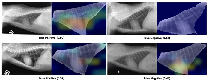
Thoracic radiograph (TR) is a complementary exam widely used in small animal medicine which requires a sharp analysis to take full advantage of Radiographic Pulmonary Pattern (RPP). Although promising advances have been made in deep learning for veterinary imaging, the development of a Convolutional Neural Networks (CNN) to detect specifically RPP from feline TR images has not been investigated. Here, a CNN based on ResNet50V2 and pre-trained on ImageNet is first fine-tuned on human Chest X-rays and then fine-tuned again on 500 annotated TR images from the veterinary campus of VetAgro Sup (Lyon, France). The impact of manual segmentation of TR’s intrathoracic area and enhancing contrast method on the CNN’s performances has been compared. To improve classification performances, 200 networks were trained on random shuffles of training set and validation set. A voting approach over these 200 networks trained on segmented TR images produced the best classification performances and achieved mean Accuracy, F1-Score, Specificity, Positive Predictive Value and Sensitivity of 82%, 85%, 75%, 81% and 88% respectively on the test set. Finally, the classification schemes were discussed in the light of an ensemble method of class activation maps and confirmed that the proposed approach is helpful for veterinarians.
This nice work was done by Ph.D. students Léo Dumortier (veterinarian) and Florent Guépin.
https://www.nature.com/articles/s41598-022-14993-2