Diagnosis models for cancer imaging
I have conducted two main projects in the field of lymphoma and prostate cancer imaging.
Both projects involve collaboration with clinicians who participate at the different steps of the CAD prototyping, sharing their clinical expertise during the definition of the technical specifications, providing annotated clinical imaging datasets as well as conducting the final diagnostic performance evaluation of the CAD prototype.
Prostate cancer detection and characterization based on multiparametric MR imaging
The joint analysis of images generated from different sequences of magnetic resonance imaging (multi-parametric MRI or mp-MRI) has been shown to allow non-invasive discrimination and localization of prostate cancer (Pca) which may open the way to targeted biopsy or active screening of patients with non aggressive cancer lesions.
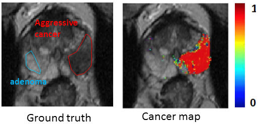
We design computer-aided diagnosis(CAD) systems for voxel- or region-based characterization of aggressive cancer in the prostate peripheral zone based on mp-MRI (including T2-weighted, diffusion-weighted and dynamic contrast enhanced MRI). Different configurations are evaluated based on the combination of supervised classification algorithm (mainly SVM) with different types of features. These features are either derived from the grey-level MR images including first-order statistics, texture parameters, or pharmacokinetic parameters derived from the dynamic data, or extracted from more advanced processing including dictionary learning. The main publications on this topic are [Niaf, PMB 2012], [Niaf, Radiol 2014], [Lehaire, ICIP 2014]
This project was funded by two grants from the french national institute for research on cancer (INCa): Cartographix (2011-14) and LYRIC (2012-17)
.
Some more details can be found here
Staging of lymphoma patients based on texture and clinical features extracted from PET and CT data
We proposed a novel CAD system that performs the automatic discrimination of cancerous from benign lesions in FDG PET/CT imaging of lymphoma patients. The optimal configuration, designed to analyse preselected suspicious areas of interest, consisted of a SVM classifier combined with a subset of 12 discriminant PET and CT features derived from the clinical practice and from more sophisticated texture analysis. An original feature selection method based on combining different filter methods was shown to outperform reference algorithms such as PCA or Genetic algorithm. The main publication on this topic is [Lartizien, IEEE JBHI 2014]