Diagnosis models for brain pathology imaging
CAD system for neuroimaging
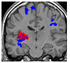 The analysis of neuroimaging data (MRI, PET..) is increasingly used in the pre-surgical work-up of patients suffering from drug resistant epilepsy. However, the detection of epilepsy lesions is very challenging as they are very heterogeneous in terms of type, size and location. We initiated a project with expert epileptologists from the Lyon Neurological Hospital to design a diagnosis model for the challenging detection of MRI negative lesions (meaning that these lesions were not visually detected by experienced neuroradiologists).
In [El Azami, Plos One 2016], we proposed to treat the epilepsy lesion detection in brain magnetic resonance (MR) images as an outlier detection problem based on the methodology developed here]. This first model combined manual engineered features to one-class SVM.
In a recent study, we proposed a novel deep unsupervised representation model based on Siamese autoencoders as an alternative to the manual feature extraction step [Alaverdyan-2018- MIDL]. This system allowed achieving sensitivity of 62% on MRI-negative lesions.
Some more details can be found here