
 French
 English
The heart is a complex organ that performs the essential function of circulating blood in the human body. This function is crucial to life, and heart disease remains a fundamental cause of death in industrialized countries. The development of diagnostic tools or therapeutic methods requires a detailed understanding of the heart’s physiology: the motion/deformation of the muscle, hemodynamics in the various cavities, electrical activation, etc. Moreover, since the heart consists of muscle fibers, it is also relevant to image the local fibrous structure of the tissue as finely as possible to establish a link between this local structure and heart function and, more generally, with the development of various pathologies.
Based on MRI imaging of water diffusion in tissue, CREATIS is a leader in cardiac fiber imaging. This imaging type is very complex, primarily because of the rapid, significant motion of the heart during MRI acquisition. In addition, thanks to the emergence of ultrafast plane-wave ultrasound imaging, the first technique for imaging tissue structure by ultrasound has recently been developed [1]. Ultrasound has many advantages over MRI, including its much lower cost, portability, and, for our application, its high acquisition speed, particularly in ultrafast imaging.
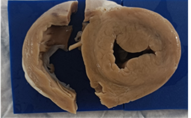
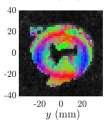
As part of the laboratory work [2-3], the technique was developed and validated across various experimental environments, including multiplexed US arrays. Based on specific signal-processing developments and new geometric considerations, the US technique appears mature for comparison with other imaging modalities and for evaluating its impact during infarction. In this context, a tomographic acquisition has already been conducted in Creatis to acquire the full morphological and fiber information of a heart slice, to, in the longer term, be able to compare such imaging to DTI, as illustrated in Fig. 1. However, the exploitation of such an acquisition pipeline is not conducted in the laboratory. Both the volume intensity and the local orientation must be extracted. To conduct such exploitation, an implicit neural representation is a potential machine learning tool, as it can enable a powerful self-supervised interpolation strategy. It has been used in medical imaging [4] and in the team [5], and similar developments have also been proposed in photoacoustic imaging [6]. Such processing seems to be an excellent candidate for our objectives.
The objectives of this master thesis project are then:
Test database construction: Using MUST simulation tools, a couple of simple geometries (points, hyper- or hypo-echoic cysts…) can be generated in a tomographic view. The simulation could be constructed in 2D or 3D, depending on the time required to run it. In addition, the already acquired dataset will be added, along with the additional information, which has already been evaluated for local orientation.
Intensity reconstruction: Using the tomographic dataset, a first effort will be made to reconstruct the intensity of the 2D/3D field-of-view using an implicit neural network. To conduct such work, the literature on such networks needs to be reviewed first and then developed in relation to the proposed database. Depending on the results, additional considerations, such as changes in attenuation or sound speed, may be warranted.
Orientation reconstruction: If the intensity reconstruction in both simulation and experiments is satisfactory, a similar strategy will be considered for local orientation estimation. In this case, no ultrasound simulation is required to generate the orientation map as proposed in Fig. 1(b). Indeed, only geometric considerations will be considered to create such maps in a first approach. However, these specific steps must be conducted in 3D, as the fibers exhibit out-of-plane orientation. The difficulty of this part consists in the fact that in each elementary volume/slice, the orientation is computed inside a local coordinate system that must be changed when evaluated in the global coordinate system.
 |  |
(a) | (b) |
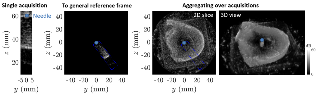 | |
Fig. 1: Current tomographic development for multimodal anisotropy imaging with (a) the heart sample extracted after animal experiments and (b) a typical DTI slice with Helix angle. (c) Illustration of the tomography test conducted in Creatis. A single volume is localized inside a general reference frame. Then, 2D or 3D morphologies are extracted. | |
General information
Profile: Student from a top engineering school or master’s degree
Technical skills: Deep learning, computer science, Python.
Soft skills: Ability to work independently, initiative, versatility, curiosity, good written and oral communication skills.
Start and duration of placement: February/March 2027 for 6 months.
Workplace: Creatis, Bat Léonard de Vinci, Campus La Doua.
How to apply
Send CV + cover letter + M1/M2 or engineering school transcripts to:
François Varray, Professor, francois.varray@creatis.insa-lyon.fr
Adrian Basarab, Professor, adrian.basarab@creatis.insa-lyon.fr
References
[1] C. Papadacci, V. Finel, J. Provost, O. Villemain, P. Bruneval, J.-L. Gennisson, M. Tanter, M. Fink, and M. Pernot, “Imaging the dynamics of cardiac fiber orientation in vivo using 3D Ultrasound Backscatter Tensor Imaging”, in Scientific Reports, 7, no. 830, 2017.
[2] R. Dumas, B. Pialot, and F. Varray. “Complete 3D anisotropy measurement with coherence imaging”, in IEEE International Ultrasonics Symposium, 2023.
[3] M. Dodel, A. Basarab, and F. Varray. “Estimating fiber orientation in ultrasound imaging through correlation and multivariate Granger causality”, in Computers in Biology and Medicine, 198(111223), 2025.
[4] A. Molaei, A. Aminimehr, A. Tavakoli, A. Kazerouni, B. Azad, A. Azad, and D. Merhof. “Implicit Neural Representation in Medical Imaging: A Comparative Survey”, in Proceedings of the IEEE/CVF International Conference on Computer Vision (ICCV) Workshops, 2023, pp. 2381-2391.
[5] F. Gaits, F. Vidal, A. Basarab, and N. Mellado, “Efficient Neural Reconstruction for Freehand 3-D Ultrasound Imaging and Visualization in Augmented Reality,” in IEEE Access, vol. 12, pp. 166253-166267, 2024.
[6] S. Huang, B. Yao, S. Cui, H. Dai, Q. Wu, Y. Xiao, F. Gao, J. Yu, Y. Zhang, X. Cai, “Implicit neural representation for sparse-view photoacoustic computed tomography”, in Ultrasonics, 7(164):107991, 2026.