
 French
 English
Context
Magnetic resonance imaging targets in an overwhelming proportion Hydrogen-1 nuclei. However other nuclei e.g. Phosphorous-31 or Hydrogen-2 also called Deuterium can both be used in magnetic metabolic applications. In the case of Deuterium, its very low endogenous concentration (0.0115%) makes it mandatory to inject Deuterium labelled agents and resort to spectroscopic imaging methods [1,2]. Using Deuterium labelled glucose [6,6′-2H2] NMR spectroscopy, it is possible to study glycolysis of tumorous cells [3] enabling more specific evaluation of the aggressivity of tumors and enhancing patient care [4]. Other medical applications include diabetes or evaluation of tissue perfusion displaying the wide potential of Deuterium metabolic imaging (DMI).
However, the low gyromagnetic ratio of Deuterium nuclei coupled with its low abundance and concentration makes the use of ultra-high magnetic field scanners and dedicated, highly sensitive Deuterium coils are mandatory for all DMI applications. This project at 11.7T aligns with the development of a new research topic within the MAGICS team of CREATIS with ongoing collaborations with research teams from the Netherlands and Japan. Moreover, with the future arrival of a clinical 7T system with x-nuclei capacities on the Lyon hospital site, this research internship will pave the way for future translational research. The intern will join the MAGICS team of CREATIS composed of experts from various fields (NMR physics, electronics, computer science, and medicine), in partnership with the PILoT imaging platform.
Internship program
During an NMR experiment, a radiofrequency transmit coil creates a ‘homogeneous’ B1 field within the studied medium flipping the magnetization into the transverse plane thus allowing its measurement. This measurement is often carried out using a second coil (receive coil) placed close to the region of interest. Such a configuration benefits both from the homogeneity of the transmitted field and from the increased sensitivity of the receiving sensor. Initial work has enabled the realization of a ²H receive coil which was interfaced on the 11.7T MRI system with first comparative images acquired as shown:
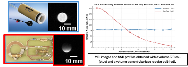
The objective of this internship is to improve the design of the surface coil and characterize its performance for Deuterium Metabolic Imaging. The intern will be responsible for the following tasks:
• Design and optimize the receive coil with active decoupling capacities: perform electronic and electromagnetic simulations, design a PCB layout, solder the components, and verify impedance matching using a VNA.
• Design Deuterium phantoms: propose a simplified model of brain tumors.
• Test the receive coil on the 11.7 T MRI system, develop a standardized experimental plan, and quantify its performance in comparison with other elements (such as a volume transmit/receive coil) available on the platform.
Required skills
• Knowledge in medical imaging
• Analog and radiofrequency electronics
• Programming
• Knowledge in Multiphysics and electronics simulation (COMSOL/LTSPICE)
Contacts
Paul NOBRE : paul.nobre@creatis.insa-lyon.fr
Kevin TSE VE KOON : kevin.tsevekoon@creatis.univ-lyon1.fr
References
[1] De Feyter et al., Deuterium metabolic imaging (DMI) for MRI-based 3D mapping of metabolism in vivo.
[2] Mahar et al., HDO production from [2H7] glucose Quantitatively Identifies Warburg Metabolism.
[3] Polvoy et al., « Deuterium Metabolic Imaging—Rediscovery of a Spectroscopic Tool ».
[4] Cember et al., « Integrating 1H MRS and Deuterium Labeled Glucose for Mapping the Dynamics of Neural Metabolism in Humans ».