The precise assessment of left ventricular filling remains an unsolved problem in diagnostic cardiology. A quantitative analysis of intracardiac blood flow could change this situation.
A large series of recent clinical studies shows that quantification of the intraventricular blood flow could lead to early diagnosis of heart failure. Intracardiac blood flow can be quantified non-invasively by cardiac magnetic resonance (CMR). Magnetic resonance velocimetry, however, is not used clinically because of its substandard cost-benefit. Another method, called echo-PIV (“echographic particle image velocimetry”) and better suited for clinical applications, is based on contrast agent echocardiography. Its main obstacle remains the intravenous administration of microbubbles, a costly and time-consuming procedure in a clinical context.
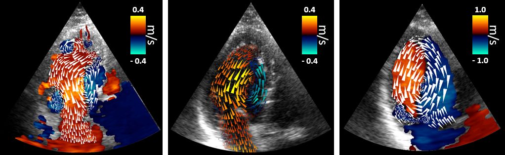
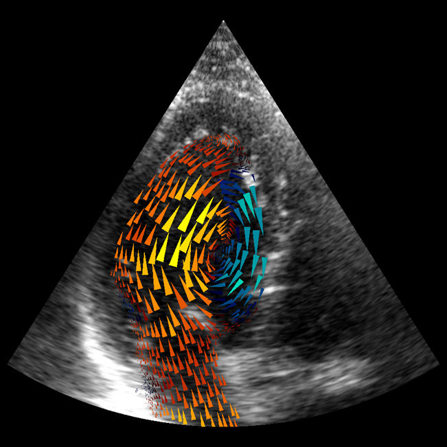
The technique of intraventricular Vector Flow Mapping (iVFM) by conventional Doppler echocardiography has thus been introduced. The original algorithm [IEEE Trans Med Imaging, 2010] has been implemented in Hitachi scanners. We recently introduced a new algorithm based on a robust regularization approach with automatic model selection [Phys Med Biol, 2017]. This new version, based on a robust regularization approach with an automatic model selection, has the advantage of being transferable to 4-D (3-D + time). In light of the success of ultrasound-derived iVFM (see articles in Google Scholar), we are extending the iVFM modality to 4-D. We expect that it will rival 4-D-CMR in clinical intracardiac velocimetry.