(E. Brusseau O. Basset)
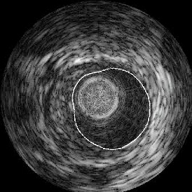
We focused on the segmentation of anatomical structures in ultrasound images. An initial approach using an active contour dedicated to the segmentation of the endoluminal contour on endovascular ultrasound images of coronary arteries was developed. This technique is based on a priori knowledge of the distribution statistics of the intensity of the image. The algorithm identifies the different Rayleigh distributions associated with each region in order to implement a Bayesian segmentation [BRUS-04b, IEEE TMI].
In relation with the Volumic imaging theme, we have also demonstrated the advantages of a multi-parametric, multi-resolution approach to the segmentation of ultrasound data [BOUK-03, Pattern Recogn Lett]. The parametric images produced make up the local measurement characteristic of the statistic distribution of ultrasound data. Estimators for the parameters for K-distributions and Nakagami distributions have been implemented to extract the information regarding the local density of the scatterers, their size and the backscattering properties of the medium [DAVI-05b, Ultrasonics].