- Participants
L. Boussel, P. Clarysse, P. Douek, M. Sigovan (Team 1)
- National and international collaborations
R. Botnar, C. Prieto, KCL (UK)
V. Rayz, Purdue University (USA)
- Question
What combination of mechanical, morphological, and physiological parameters is related to rapid progression of vascular diseases? How to accurately quantify and analyze blood flow and flow related biomarkers in different vascular territories?
- Objective
- To evaluate in a joint manner the mechanical, morphological, and physiological parameters related to vascular disease progression to provide a better understanding of their roles and interactions and to improve diagnosis and predict patient outcome.
- To develop 7D phase contrast MRI methods for the accurate quantification of blood flow.
- To develop post-processing methods for quantification of flow related biomarkers.
- Method
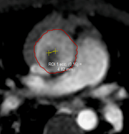
Our approach is based on the combination of 3D radial k-space sampling trajectory with respiratory self-gating and motion correction to obtain 100% scan efficiency and isotropic millimeter resolution. Segmentation, post-processing, and numerical modeling methods are developed and performed on data acquired in longitudinal studies on patients presenting with vascular disease (atherosclerosis, aneurysms, dissections) using MRI and hybrid PET/MRI systems. Hemodynamic biomarkers such as wall shear stress, flow displacement, are computed and correlated to disease progression (Figure 1).
- Illustration
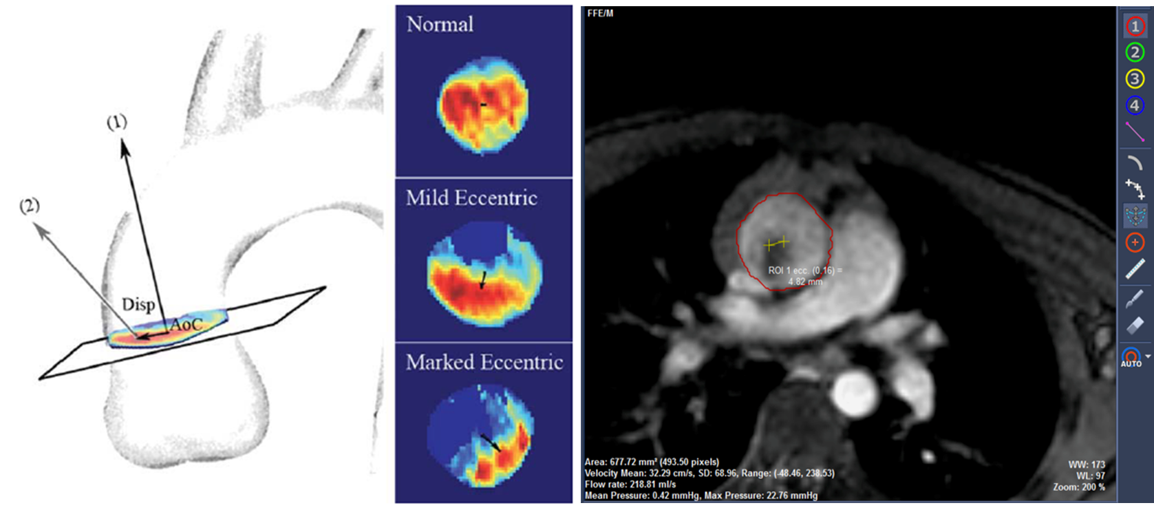
Figure 1. Left: computation of the flow displacement parameter at the level of the ascending aorta [Sigovan et al, JMRI 2011]. We developed in 2D and 3D and demonstrated the predictive value of this parameter for the progression of thoracic aortic aneurysms; Right: implementation of flow displacement parameter in commercial software of cardio-vascular image analysis dedicated to radiologists (Medis, Leiden, The Netherlands)
- Bibliographic references
- Journals
M Sigovan, P Dyverfeldt, J Wrenn, EE Tseng, D Saloner, MD Hope. Extended 3D approach for quantification of abnormal ascending aortic flow. Magn Reson Imaging. 2015 Jun;33(5):695-700.
MD Hope, M Sigovan, SJ Wrenn, D Saloner, P Dyverfeldt. MRI hemodynamic markers of progressive bicuspid aortic valve-related aortic disease. J Magn Reson Imaging 2014 Jul;40(1):140-5
M. Sigovan, V. Rayz, W. Gasper, H. Alley, D. Saloner. “Vascular remodeling in Autogenous Arterio-Venous Fistulas by MRI and CFD”. Annals of Biomedical Engineering, 2013 Apr;41(4):657-68.
- Conferences
M. Sigovan, M. Roy, V. Rayz, P. Douek, L. Boussel. “Gadolinium enhancement vs WSS and Oscillatory shear Index (OSI) in human carotid atherosclerosis by MRI and CFD: A preliminary Study”. The Magnetic Resonance Angio Club, Rome, Italy, September 16-19, 2014.
M. Sigovan, L. Boussel, A. Millon, P. Douek, E. Canet-Soulas. “Low WSS drives eccentric plaque formation in an animal model of induced atherosclerosis”. The Magnetic Resonance Angio Club, New York, United States, August 20-23, 2013.