Key words: MR sequence & post-processing development, muscle dynamics, longitudinal & functional variations studies
Scientific and application contexts
Characterising and predicting functional changes and localised mechanical risks in skeletal muscle using imaging is a major challenge. The current challenge is to enable a better understanding of individual anatomical and pathophysiological data for optimised training planning and injury prevention, whether for sedentary individuals resuming physical activity or for monitoring high-level athletes in a preventive sports medicine approach.
Recent innovative developments in MRI make it possible to envisage non-invasive, in-depth exploration of functional alterations in skeletal muscle, placing imaging at the centre of clinical investigations. Image analysis and processing tools are essential assets for clinical research teams, both from an innovation perspective and in terms of their value within the medical and scientific community. The Saint-Etienne University Hospital (CHUSE) and CREATIS have a full set of Ergospect ergometric modules that enable controlled muscle exercises to be performed in an MRI scanner. It also has the Cima X research MRI scanner from Siemens Healthcare, equipped with the most powerful gradient system available in clinical practice.
Databases and prior knowledge
The project is based on MRI databases in the fields of sport (MUST project, HAMMER project, upcoming ‘0 to 100’ project), nutrition (COPHYAM cohort (normal, extreme thinness and anorexia), GENESIS (therapeutic fasting nutritional intervention (3 stages)), ANIMATION), or dedicated to the study of post-COVID or post-MS fatigue (FatPostRéa (two stages: upon discharge from intensive care and post-functional rehabilitation), FatPostSEP (multiple sclerosis)). Two studies have investigated skeletal muscle recovery after extreme exercise by tracking water distribution within exercising muscles using advanced MRI imaging techniques [Froeling 2015, Gilles 2016]. It has been shown [Saugy 2013] that during extreme exercise such as 24-hour running, the loss of strength in the knee extensor muscles averaged 40% and that three-quarters of this fatigue could be explained by central fatigue and only 25% by alterations in contractile structures. The aetiology of fatigue is complex, but it is accepted that its extent and origins are specific to the exercise performed, in terms of duration and intensity, the subject's morphotype, but also depending on the geographical profile of the race, involving variations in contraction modes (eccentric versus concentric). In the case of ultra-marathons in the mountains, it is very likely that muscle damage is even more severe due to the existence of a marked and prolonged eccentric component during the downhill sections [Schütz 2013].
Objectives
The subject of the post-doctoral position will be:
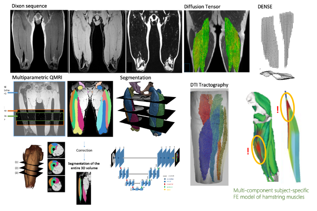
i) to improve the reliability of static and dynamic acquisition protocols in diffusion MRI of skeletal muscle, to build a robust and automatic processing chain to obtain metrics and data derived from tractography and fibre strain to feed into numerical simulations (carried out by another part of the project). More specifically, this will involve defining protocols for acquiring MRI data of muscles at rest and during exercise (similar to techniques used to explore the heart muscle) in order to ensure robustness and reliability. MRI is also capable of objectively assessing the inflammatory phenomena caused by intense physical exercise, which can impact post-exercise recovery. This information should make it possible to personalise and build a new model of human tissue stress in order to improve recovery and prevent muscle damage. MRI is also capable of more finely characterising changes in muscle composition linked to improved oxygenation, muscle metabolism and/or lipid composition. The project will therefore require innovative and advanced acquisition protocols (diffusion imaging, DENSE motion encoding imaging on the one hand, and on the other hand, data analysis and the implementation of a robust and automated pipeline [Nguyen 2018], before creating a surface or volume geometry adapted to numerical simulation tools and finally creating a numerical simulation model. Post-processing will provide multi-parametric metrics and functional information relevant to numerical simulations.
The new MRI protocol will enable:
a) a better understanding of the physiological mechanisms involved in muscle movement.
b) the study of the relevance of non-invasive indicators available in MRI on a relevant model of inflammation.
c) the acceleration of the transfer of these technologies to the clinical setting, for athletes and patients.
Research program and proposed scientific approach:
Semester 1: Familiarisation with the context: bibliography (identification of methods and teams working on similar topics), creation of a robust processing chain for the segmentation of lower limb muscle heads, pre- and post-processing. Deliverables: MRI protocols, Matlab or Python processing pipeline.
Semester 2: Deployment of imaging methods and protocols on healthy volunteers, validation, analysis of data produced and reproducibility study (longitudinal). Deliverables: Matlab or Python plug-in, slicer plug-in, applied journal with code distribution. Submission of abstracts and conferences.
Semester 3: Combination of structural, anatomical and functional information on volunteers who are athletes, injured or not. Deliverable: an approach and a database validated by experts.
Semester 4: Combination of structural and functional data produced and surface mesh constraints for numerical simulation. Co-definition of the specifications for the geometry, meshes and labels to be generated automatically. Study and management of discrepancies between expert/automatic/mesh segmentations. Sharing with other parts of the project. Deliverables: Slicer plug-in for the proposed approach. Publications.
Candidate profile
Proficiency in MRI, particularly cardiac MRI, proficiency in medical image processing and analysis, particularly in segmentation and modelling, knowledge of digital processing issues (meshing, segmentation, registration). Ability to communicate across disciplines in French and English.
Bibliography references on the subject
[Froeling 2015] “Muscle changes detected with diffusion-tensor imaging after long-distance running”. Radiology. 274(2):548-62. 2015
[Gilles 2016] “Automatic segmentation of quadriceps muscle head and volume quantification”, ISMRM 2016, Singapour,
[Jouvencel 2022] « Impact of MR sequences choice on deep learning segmentation of muscles ». In : 2022 16th IEEE International Conference on Signal Processing (ICSP). T. 1, p. 420-425.
[Naegel 2025] « Impact of Long-Term Fasting on Skeletal Muscle : Structure, Energy Metabolism and Function Using 31P/1H MRS and MRI ». In : Journal of Cachexia, Sarcopenia and Muscle 16.2, e13773.
[Nguyen 2021a] « Impact of distortion on local radiomic analysis of quadriceps based on quantitative magnetic resonance imaging data ». In : International Journal of Pharma Medicine and Biological Sciences 10.2, p. 49-54.
[Nguyen 2021b] « Quantitative magnetic resonance imaging assessment of the quadriceps changes during an extreme mountain ultramarathon ». In : Medicine & Science in Sports & Exercise 53.4, p. 869-881.
[Nguyen 2018] « Robust multi-atlas MRI segmentation with corrective learning for quantification of local quadriceps muscles inflammation changes during a longitudinal study in athletes ». In : Proc. Intl. Soc. Mag. Reson. Med. 26. Paris, France.
[Saillard 2024] « Finite element models with automatic computed tomography bone segmentation for failure load computation ». In : Scientific Reports 14.1, p. 16576.
[Saugy 2013] Alterations of Neuromuscular Function after the World's Most Challenging Mountain Ultra-Marathon. PLOS One, June 26, 2013
[Schütz 2013] Characteristics, changes and influence of body composition during a 4486 km transcontinental ultramarathon: results from the TransEurope FootRace mobile whole body MRI-project. BMC Med. 2013 May 8;11:122.
[Mazzoli 2021] Front Neurol. 15;12:608549. doi: 10.3389/fneur.2021.608549
[Verzhbinsky 2023] IEEE Trans Med Imaging. 2020 Mar;39(3):656-667. doi: 10.1109/TMI.2019.2933813.
[Naegel 2025] Impact of Long-Term Fasting on Skeletal Muscle: Structure, Energy Metabolism and Function Using 31P/1H MRS and MRI. J Cachexia Sarcopenia Muscle. 2025 Apr;16(2):e13773. doi: 10.1002/jcsm.13773.
PMID: 40211897
[Wang 2023] StrainNet: Improved Myocardial Strain Analysis of Cine MRI by Deep Learning from DENSE. Radiol Cardiothorac Imaging. 2023 May 4;5(3):e220196. doi: 10.1148/ryct.220196. eCollection 2023 Jun. PMID: 37404792
TEAM : CREATIS – MAGICS- CHUSE
Principal Investigator : Pr. Pierre Croisille.
Funding: Tanenbaum Institute of Science for Sports (TISS) and CHU of Saint-Etienne.
Co-investigators :Magalie Viallon, Pierre Croisille
Contacts : pierre.croisille@creatis.insa-lyon.fr ou magalie.viallon@creatis.insa-lyon.fr