Research interests
Signal and image processing for computational biomedical imaging: inverse problems applied to medical imaging, from model-based methods to machine learning. Select a topic for details.
Acquisition and image formation
Image reconstruction and restoration
Quantitative and functional imaging
Quantum mechanics-inspired signal and image processing
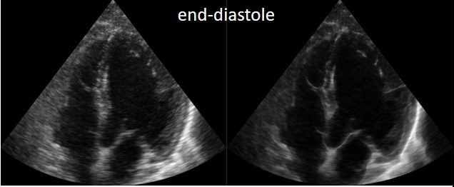
Cardiac ultrasound at end-diastole: original image from a clinical scanner (left) and enhanced image (right). Dutta et al., IEEE IUS, 2022. Context Denoising and restoration remain central to medical imaging, where noise statistics are often far from Gaussian (speckle in ultrasound, Poisson noise in CT) and where hand-crafted priors struggle to adapt to local image content.
Contribution In the PhD thesis of Sayantan Dutta , co-supervised with Denis Kouamé and Bertrand Georgeot (Laboratoire de Physique Théorique, Toulouse), we built adaptive signal and image representations from quantum mechanics: the image is treated as a potential in the Schrödinger equation, and the localization of its wave functions separates signal from noise. We extended the idea to quantum many-body interactions between patches, plugged the resulting denoisers into plug-and-play schemes for deconvolution, and unfolded them into deep networks, with applications to ultrasound despeckling, cardiac image enhancement and dental CT restoration.
Selected references S. Dutta, A. Basarab , B. Georgeot and D. Kouamé, “Quantum mechanics-based signal and image representation: application to denoising,” IEEE Open Journal of Signal Processing , vol. 2, pp. 190–206, 2021. S. Dutta, A. Basarab , B. Georgeot and D. Kouamé, “A novel image denoising algorithm using concepts of quantum many-body theory,” Signal Processing , vol. 201, 108690, 2022. S. Dutta, A. Basarab , B. Georgeot and D. Kouamé, “DIVA: deep unfolded network from quantum interactive patches for image restoration,” Pattern Recognition , vol. 155, 110676, 2024. S. Dutta, B. Georgeot, D. Kouamé, D. Garcia and A. Basarab , “Adaptive contrast enhancement of cardiac ultrasound images using a deep unfolded many-body quantum algorithm,” IEEE International Ultrasonics Symposium , 2022. Code
Fusion of MR and ultrasound images
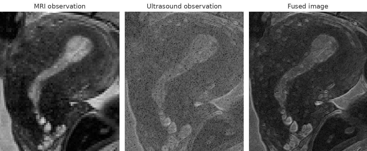
Low-resolution MRI, noisy ultrasound image simulated from a high-resolution MRI, and the image fused by our method, which combines the contrast of MRI with the resolution of ultrasound. El Mansouri et al., IEEE TIP, 2020. Context Endometriosis surgery relies on MRI for its wide field of view and good contrast, and on ultrasound for its high spatial resolution, but the two modalities are acquired and read separately.
Contribution In collaboration with Jean-Yves Tourneret, we proposed fusion methods that combine the contrast and signal-to-noise ratio of MRI with the resolution of ultrasound in a single image. The fusion is formulated as an inverse problem with modality-specific observation models (super-resolution for MRI, despeckling for ultrasound) linked by a polynomial relation between gray levels, and was later extended to joint registration and fusion of 3D MRI and 2D ultrasound.
Selected references O. El Mansouri, F. Vidal, A. Basarab , P. Payoux, D. Kouamé and J.-Y. Tourneret, “Fusion of magnetic resonance and ultrasound images for endometriosis detection,” IEEE Transactions on Image Processing , vol. 29, pp. 5324–5335, 2020. Y. El Bennioui, F. Vidal, A. Basarab and J.-Y. Tourneret, “Joint registration and fusion of 3D magnetic resonance and 2D ultrasound images for endometriosis surgery,” EUSIPCO , 2023. Y. El Bennioui, A. Halimi, A. Basarab and J.-Y. Tourneret, “Fusion of magnetic resonance and ultrasound images using guided filtering: application to endometriosis surgery,” EUSIPCO , 2024. Code
Artifact quantification in lung ultrasound
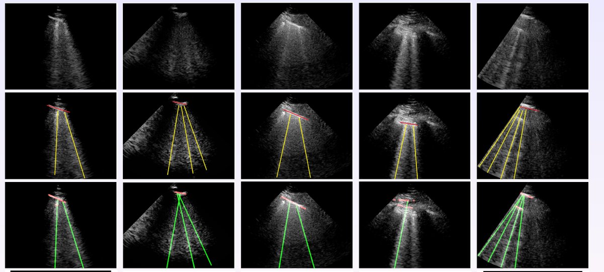
Original images (top), ground-truth B-lines (yellow) and lines detected by our method (green). Karakuş et al., IEEE TUFFC, 2020. Context Lung ultrasound is cheap, portable and radiation-free, and was widely used during the COVID-19 pandemic. Its diagnosis relies on artifacts such as B-lines, whose manual counting is time-consuming and operator-dependent.
Contribution We formulated line detection as an inverse problem in the Radon domain, with a non-convex, Cauchy-based penalty that promotes a small number of lines, and evaluated it on COVID-19 patients.
Selected references O. Karakuş, N. Anantrasirichai, A. Aguersif, S. Silva, A. Basarab and A. Achim, “Detection of line artefacts in lung ultrasound images of COVID-19 patients via non-convex regularization,” IEEE Transactions on Ultrasonics, Ferroelectrics and Frequency Control , vol. 67, no. 11, pp. 2218–2229, 2020. Code
Ultrasound image restoration: deconvolution, super-resolution and diffusion models
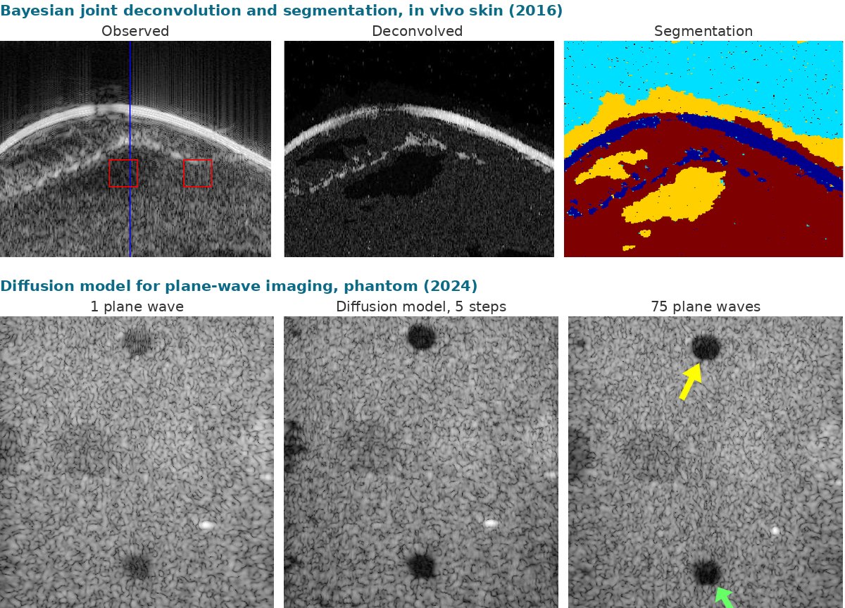
Top: joint deconvolution and segmentation of an in vivo skin image (Zhao et al., IEEE TIP, 2016). Bottom: phantom imaged with a single plane wave, denoised by our diffusion model in 5 steps, and imaged with 75 compounded plane waves (Asgariandehkordi et al., IEEE TUFFC, 2024). Context The limited bandwidth of ultrasound transducers and wave propagation in tissue degrade spatial resolution and contrast. Under the first-order Born approximation and weak scattering, these degradations follow a linear model relating the tissue reflectivity function to the RF data. In plane-wave imaging, a single emission gives very high frame rates but noisy, low-contrast images, and compounding many angles restores quality at the cost of frame rate.
Contribution With Denis Kouamé and Jean-Yves Tourneret, our group has long worked on inverting this image formation model to recover images closer to the tissue than native ultrasound images: Bayesian joint deconvolution and segmentation with generalized Gaussian priors, fast analytical solutions for single-image super-resolution, and deconvolution exploiting both fundamental and harmonic images. With Hassan Rivaz (Concordia University), we extended this line in two directions. On the model-based side, we cast beamforming as an inverse problem regularized by denoisers and unified beamforming and deconvolution in a single problem. On the learning side, we adapted denoising diffusion probabilistic models to plane-wave data: the reverse process starts from a single plane-wave image instead of pure noise and reaches the quality of compounded images in a few deterministic steps.
Selected references N. Zhao, A. Basarab , D. Kouamé and J.-Y. Tourneret, “Joint segmentation and deconvolution of ultrasound images using a hierarchical Bayesian model based on generalized Gaussian priors ,” IEEE Transactions on Image Processing , vol. 25, no. 8, pp. 3736–3750, 2016. N. Zhao, Q. Wei, A. Basarab , N. Dobigeon, D. Kouamé and J.-Y. Tourneret, “Fast single image super-resolution using a new analytical solution for ℓ2–ℓ2 problems ,” IEEE Transactions on Image Processing , vol. 25, no. 8, pp. 3683–3697, 2016. M. Hourani, A. Basarab , D. Kouamé and J.-Y. Tourneret, “Ultrasound image deconvolution using fundamental and harmonic images,” IEEE Transactions on Ultrasonics, Ferroelectrics and Frequency Control , vol. 68, no. 4, pp. 993–1006, 2021. S. Goudarzi, A. Basarab and H. Rivaz, “Inverse problem of ultrasound beamforming with denoising-based regularized solutions,” IEEE Transactions on Ultrasonics, Ferroelectrics and Frequency Control , vol. 69, no. 10, pp. 2906–2916, 2022. S. Goudarzi, A. Basarab and H. Rivaz, “A unifying approach to inverse problems of ultrasound beamforming and deconvolution,” IEEE Transactions on Computational Imaging , vol. 9, pp. 197–209, 2023. H. Asgariandehkordi, S. Goudarzi, M. Sharifzadeh, A. Basarab and H. Rivaz, “Denoising plane wave ultrasound images using diffusion probabilistic models ,” IEEE Transactions on Ultrasonics, Ferroelectrics and Frequency Control , vol. 71, no. 11, pp. 1526–1539, 2024. Code
Compressed sensing and sparsity in ultrasound
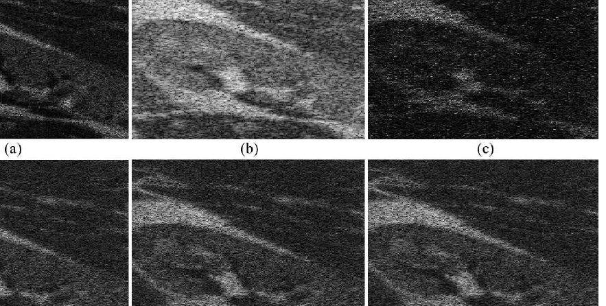
Compressive deconvolution of a simulated kidney image. Chen, Basarab and Kouamé, IEEE TMI, 2016. Context New acquisition modes such as ultrafast imaging, based on plane-wave emissions, and 3D imaging either lower image quality or produce very large amounts of data to collect and process.
Contribution In 2010, our group was among the first to use compressed sensing to reduce the data volume in ultrasound imaging. This pioneering work was pursued by several groups and established sparsity as an important alternative to classical Gaussian signal processing. We then combined compressed sensing and deconvolution, recovering enhanced images directly from compressed measurements.
Selected references C. Quinsac, A. Basarab and D. Kouamé, “Frequency domain compressive sampling for ultrasound imaging ,” Advances in Acoustics and Vibration , vol. 12, pp. 1–16, 2012. Z. Chen, A. Basarab and D. Kouamé, “Compressive deconvolution in medical ultrasound imaging ,” IEEE Transactions on Medical Imaging , vol. 35, no. 3, pp. 728–737, 2016. Z. Chen, A. Basarab and D. Kouamé, “Reconstruction of enhanced ultrasound images from compressed measurements using simultaneous direction method of multipliers,” IEEE Transactions on Ultrasonics, Ferroelectrics and Frequency Control , vol. 63, no. 10, pp. 1525–1534, 2016. Code
Continuous-emission ultrasound imaging
Pulse-echo: emit, then wait for the echoes listen listen listen Continuous emission: random coded waveform, never interrupted sliding windows → one line each Schematic: pulse-echo imaging alternates short pulses and listening periods, whereas continuous emission sends an uninterrupted random coded waveform decoded with sliding windows. Context In pulse-echo ultrasound, the pulse repetition frequency must stay low enough to avoid range ambiguity, which limits the frame rate; the medium is also observed only for a short time after each pulse, so fast or short-lived events can be missed.
Contribution In the PhD thesis of Axel Adam , co-supervised with Barbara Nicolas and Hervé Liebgott, we proposed continuous-emission ultrasound imaging, inspired by radar and sonar. The medium is insonified without interruption with a random coded waveform, and sliding windows on the emitted and received signals produce one image line every few samples, which raises the slow-time rate by about two orders of magnitude compared with pulse-echo imaging. We designed optimized mismatched filters and inverse-problem decoders, extended the approach from M-mode to B-mode imaging, and validated it experimentally.
Selected references A. Adam, B. Nicolas, A. Basarab and H. Liebgott, “Continuous emission ultrasound: a new paradigm to ultrafast ultrasound imaging ,” IEEE Transactions on Biomedical Engineering , vol. 72, no. 1, pp. 388–400, 2025. A. Adam, A. Basarab , H. Liebgott and B. Nicolas, “An optimized mismatched filter for continuous emission ultrasound imaging,” EUSIPCO , 2024. A. Adam, H. Liebgott, B. Nicolas and A. Basarab , “High-framerate B-mode ultrasound imaging using continuous emission,” IEEE International Symposium on Biomedical Imaging (ISBI) , 2025. M. Tamraoui, A. Adam, A. Basarab , B. Nicolas and H. Liebgott, “Experimental validation of continuous emission ultrafast ultrasound imaging,” IEEE International Ultrasonics Symposium , 2025. Code No public code available yet.
Tissue motion estimation
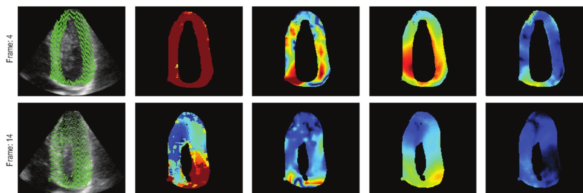
Motion fields and error maps in echocardiography for several methods, ours on the right. Ouzir et al., IEEE TIP, 2018. Context Estimating tissue motion from image sequences is key to assessing cardiac function and to elastography, but ultrasound speckle decorrelation and noise make it difficult.
Contribution Since my PhD, I have developed several motion estimation methods. For many years, their main originality was the use of spatial phase images (monogenic signal, transverse oscillations) instead of native ultrasound images. More recently, we showed the value of sparse representations and dictionary learning for cardiac motion estimation.
Selected references M. Alessandrini, A. Basarab , H. Liebgott and O. Bernard, “Myocardial motion estimation from medical images using the monogenic signal ,” IEEE Transactions on Image Processing , vol. 22, no. 3, pp. 1084–1095, 2013. N. Ouzir, A. Basarab , H. Liebgott, B. Harbaoui and J.-Y. Tourneret, “Motion estimation in echocardiography using sparse representation and dictionary learning,” IEEE Transactions on Image Processing , vol. 27, no. 1, pp. 64–77, 2018. N. Ouzir, A. Basarab , O. Lairez and J.-Y. Tourneret, “Robust optical flow estimation in cardiac ultrasound images using a sparse representation ,” IEEE Transactions on Medical Imaging , vol. 38, no. 3, pp. 741–752, 2019. Code
Dental CT super-resolution and segmentation
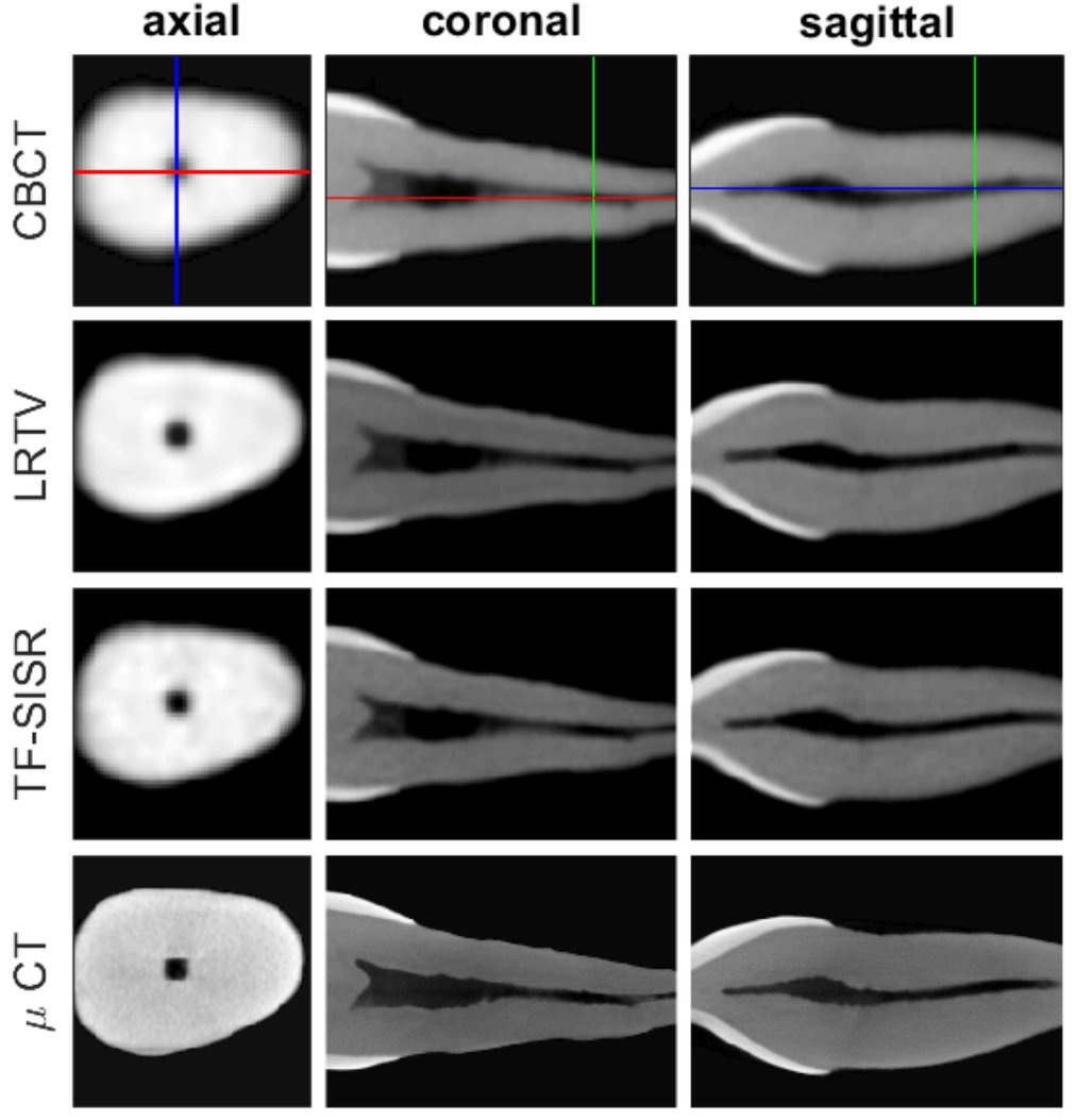
Tooth imaged by CBCT, super-resolved by two methods, and by micro-CT for reference. Hatvani et al., IEEE TMI, 2019. Context In endodontics, root canals must be precisely located and measured, but clinical cone-beam CT has a limited resolution compared with micro-CT, which cannot be used in patients.
Contribution We proposed super-resolution methods for 3D dental CBCT, based on tensor factorization and on deep learning, and evaluated 3D segmentation of root canals against micro-CT references.
Selected references J. Hatvani, A. Basarab , J.-Y. Tourneret, M. Gyöngy and D. Kouamé, “A tensor factorization method for 3D super-resolution with application to dental CT,” IEEE Transactions on Medical Imaging , vol. 38, no. 6, pp. 1524–1531, 2019. J. Hatvani, A. Horváth, J. Michetti, A. Basarab , D. Kouamé and M. Gyöngy, “Deep learning-based super-resolution applied to dental computed tomography ,” IEEE Transactions on Radiation and Plasma Medical Sciences , vol. 3, no. 2, pp. 120–128, 2019. J. Michetti, A. Basarab , F. Diemer and D. Kouamé, “Comparison of an adaptive local thresholding method on CBCT and µCT endodontic images ,” Physics in Medicine and Biology , vol. 63, pp. 1–10, 2018. Code
Augmented reality and freehand 3D ultrasound
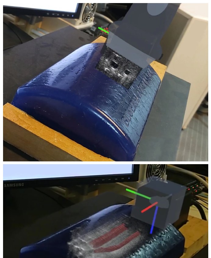
Augmented reality display on a phantom: the current ultrasound slice shown in place (top) and the reconstructed 3D volume with the tracked probe (bottom). Gaits et al., IEEE Access, 2024. Context Freehand 2D ultrasound is cheap and real-time, but the operator must mentally reconstruct 3D anatomy from moving slices. Showing images directly in the patient space, and turning freehand sweeps into 3D volumes, could make ultrasound easier to use and to teach.
Contribution In the PhD thesis of François Gaits , co-supervised with Nicolas Mellado, and in collaboration with Fabien Vidal, physician, we built tools to simulate, reconstruct and visualize freehand ultrasound in augmented reality: an efficient 3D scatterer sampling for realistic freehand simulation, neural implicit representations for 3D volume reconstruction from tracked 2D frames, and a real-time framework that streams experimental and simulated images to an augmented reality headset. These developments are gathered in the T-Play platform. In the ongoing PhD thesis of Ziyi Chen, co-supervised with Shaifali Parashar, we develop deformation-aware simulation of handheld ultrasound.
Selected references F. Gaits, N. Mellado, G. Bouyjou, D. Garcia and A. Basarab , “Efficient stratified 3D scatterer sampling for freehand ultrasound simulation,” IEEE Transactions on Ultrasonics, Ferroelectrics and Frequency Control , vol. 71, no. 1, pp. 127–140, 2024. F. Gaits, F. Vidal, A. Basarab and N. Mellado, “Efficient neural reconstruction for freehand 3-D ultrasound imaging and visualization in augmented reality,” IEEE Access , vol. 12, pp. 166253–166267, 2024. F. Gaits, G. Bouyjou, E. Boni, A. Ramalli, P. Tortoli, A. Basarab et al., “A framework for real-time visualization of experimental and simulated ultrasound images in augmented reality,” IEEE International Ultrasonics Symposium , 2024. Z. Chen, S. Parashar and A. Basarab , “Deformation-aware simulator for handheld ultrasound imaging,” IEEE International Symposium on Biomedical Imaging (ISBI) , 2026. Code
Passive cavitation imaging
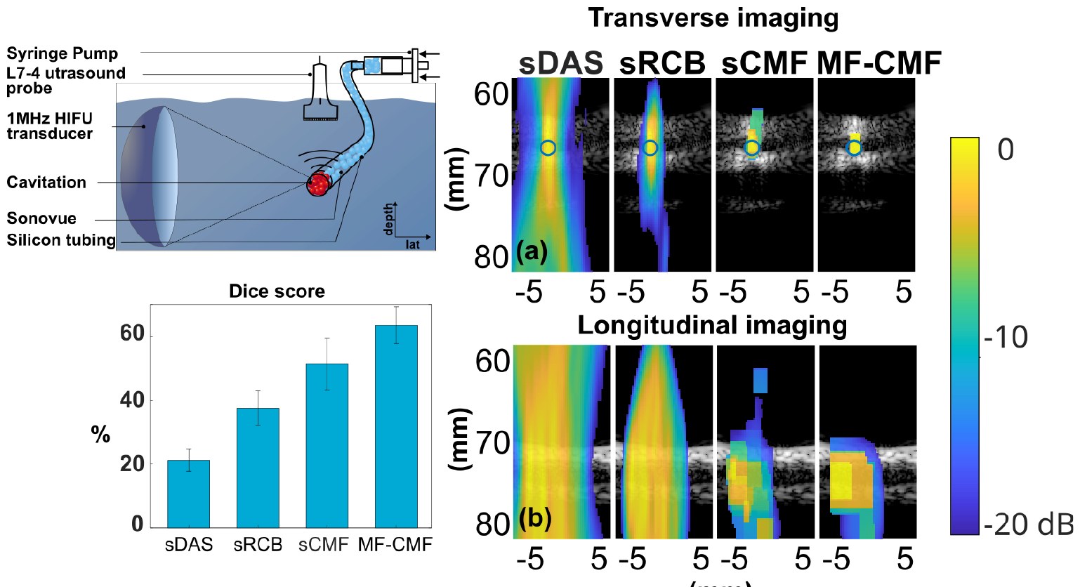
Cavitation induced by focused ultrasound in a tube: setup, Dice scores and cavitation maps for delay-and-sum, robust Capon, cross-spectral matrix fitting and its multi-frequency version. Lachambre et al., IEEE IUS, 2025. Context Cavitation induced by high-intensity focused ultrasound must be monitored for therapies such as sonoporation. Passive acoustic mapping localizes cavitation from the signals it emits, but conventional beamformers such as delay-and-sum or robust Capon have a poor axial resolution and produce artifacts.
Contribution In the PhD thesis of Célestine Lachambre , co-supervised with Bruno Gilles, and in collaboration with Barbara Nicolas, François Varray and Jean-Christophe Béra, we formulated passive cavitation imaging as an inverse problem on cross-spectral matrices, with sparsity-promoting and elastic-net regularization, then combined several frequencies and introduced learned priors. With Tatiana Gelvez during her postdoc, we proposed time-domain linear models and efficient convolutional forward models for passive acoustic mapping and the temporal monitoring of cavitation activity.
Selected references C. Lachambre, A. Basarab , J.-C. Béra, B. Nicolas, F. Varray and B. Gilles, “An inverse method using cross spectral matrix fitting for passive cavitation imaging,” IEEE Transactions on Ultrasonics, Ferroelectrics and Frequency Control , vol. 71, no. 8, pp. 995–1005, 2024. C. Lachambre, B. Gilles, J.-C. Béra, F. Varray, B. Nicolas and A. Basarab , “Frequency compounding cross spectral matrix fitting for passive cavitation imaging,” IEEE Transactions on Ultrasonics , 2026. T. Gelvez-Barrera, B. Nicolas, D. Kouamé, B. Gilles and A. Basarab , “Time-domain linear model-based framework for passive acoustic mapping of cavitation activity,” IEEE Transactions on Ultrasonics , 2026. C. Lachambre, B. Gilles, B. Nicolas, F. Varray, J.-C. Béra and A. Basarab , “Introduction of a learned prior to passive cavitation imaging,” IEEE International Symposium on Biomedical Imaging (ISBI) , 2026. Code No public code available yet.
Quantitative ultrasound: attenuation, backscatter and tissue anisotropy
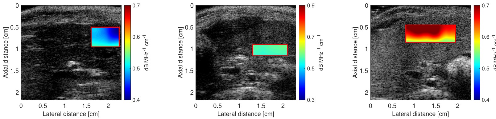
Attenuation coefficient maps in a murine model of liver steatosis, for increasing steatosis grades (left to right). Timaná et al., Scientific Reports, 2023. Context B-mode images are qualitative and depend on the scanner settings. Quantitative ultrasound estimates intrinsic tissue properties, such as the attenuation and backscatter coefficients or the orientation of fibers, which can help diagnose diseases like liver steatosis, but these estimates are noisy and have a limited spatial resolution.
Contribution With Roberto Lavarello (Pontificia Universidad Católica del Perú), we cast the estimation of attenuation and backscatter coefficients as regularized inverse problems: simultaneous estimation of both coefficients, joint multi-frequency reconstruction with total nuclear variation, regularization by denoising, and deep unfolded networks (ACS-Net, best student paper finalist at IEEE IUS 2025). In the PhD thesis of Mahshid Dodel, co-supervised with François Varray, we estimate fiber orientation in ultrasound images with correlation and multivariate Granger causality.
Selected references J. Timaná, H. Chahuara, L. Basavarajappa, A. Basarab , K. Hoyt and R. Lavarello, “Simultaneous imaging of ultrasonic backscatter and attenuation coefficients for quantitative liver steatosis assessment,” Scientific Reports , vol. 13, 2023. E. Miranda, J. Timaná, A. Basarab and R. Lavarello, “Enhancing ultrasonic attenuation images through multi-frequency coupling with total nuclear variation,” The Journal of the Acoustical Society of America , vol. 156, no. 4, pp. 2805–2815, 2024. S. Merino, A. Carrera, E. Avilés, A. Basarab and R. Lavarello, “Attenuation coefficient imaging using regularization by denoising,” IEEE Transactions on Ultrasonics , 2026. M. Dodel, A. Basarab and F. Varray, “Estimating fiber orientation in ultrasound imaging through correlation and multivariate Granger causality,” Computers in Biology and Medicine , vol. 198, 111223, 2025. Code No public code available yet.
Quantitative acoustic microscopy
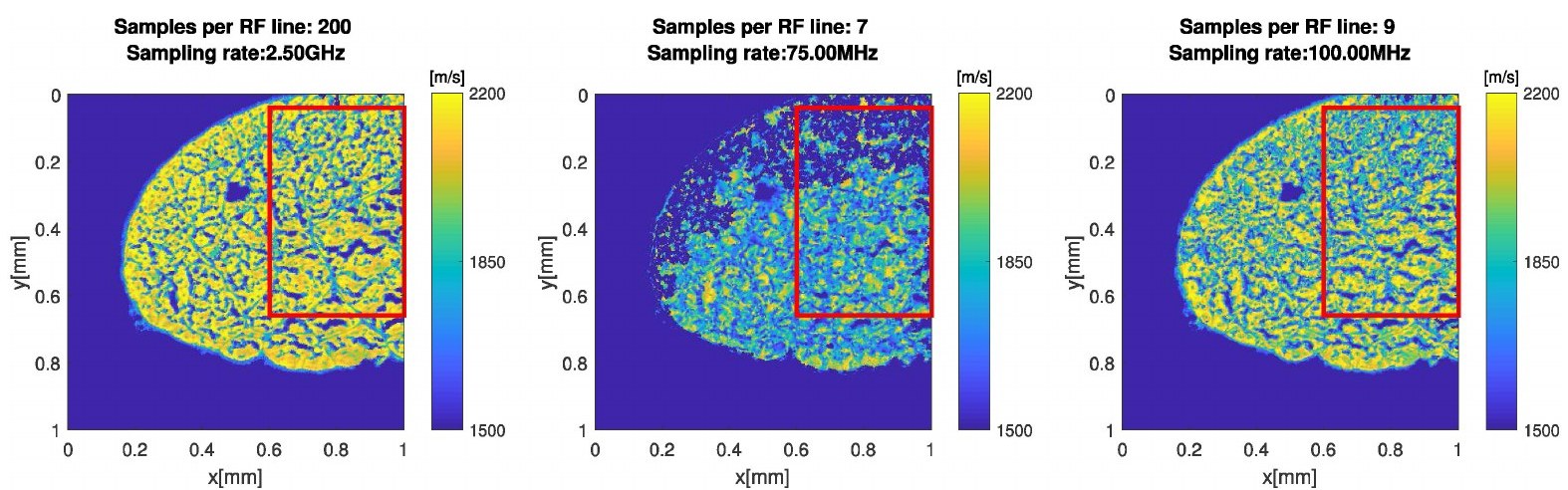
Speed-of-sound maps from fully sampled RF data (200 samples per line, left) and reconstructed from only 7 and 9 samples per line. Kim et al., IEEE TCI, 2020. Context Quantitative acoustic microscopy at very high frequencies (250 MHz and above) maps mechanical properties of thin tissue sections, such as speed of sound, acoustic impedance and attenuation, at micrometer resolution. Acquisition requires very high sampling rates and long scanning times.
Contribution In collaboration with Jonathan Mamou (Cornell University, NIH-funded project) and Alin Achim (University of Bristol), we reduced acquisition requirements while preserving image quality. In the PhD thesis of Jonghoon Kim, we used compressed sensing with approximate message passing, and models with a finite rate of innovation to reconstruct quantitative maps from RF signals sampled far below the Nyquist rate. With Lorena Leon during her postdoc, we proposed weighted Hankel spectral methods and analyzed their performance with Cramér–Rao bounds; we also unfolded message passing into deep networks.
Selected references J. Kim, J. Mamou, P. Hill, N. Canagarajah, D. Kouamé and A. Basarab , “Approximate message passing reconstruction of quantitative acoustic microscopy images ,” IEEE Transactions on Ultrasonics, Ferroelectrics and Frequency Control , vol. 65, no. 3, pp. 327–338, 2018. J.-H. Kim, J. Mamou, D. Kouamé, A. Achim and A. Basarab , “Autoregressive model-based reconstruction of quantitative acoustic maps from RF signals sampled at innovation rate,” IEEE Transactions on Computational Imaging , vol. 6, pp. 993–1006, 2020. L. Leon, J. Mamou, D. Kouamé and A. Basarab , “A weighted Hankel approach and Cramér–Rao bound analysis for quantitative acoustic microscopy imaging,” Computers in Biology and Medicine , vol. 196, 110730, 2025. O. Pappas, J. Mamou, A. Basarab , D. Kouamé and A. Achim, “Deep unfolded approximate message passing for quantitative acoustic microscopy image reconstruction,” IEEE ICASSP , 2025. Code No public code available yet.
 Passive cavitation imaging
 Augmented reality and freehand 3D ultrasound
 Compressed sensing in ultrasound
 Ultrasound image restoration
 Quantum-inspired image processing
 MR–ultrasound image fusion
 Dental CT super-resolution